Filiform papillae, light micrograph
Numéro d’image : 13673457
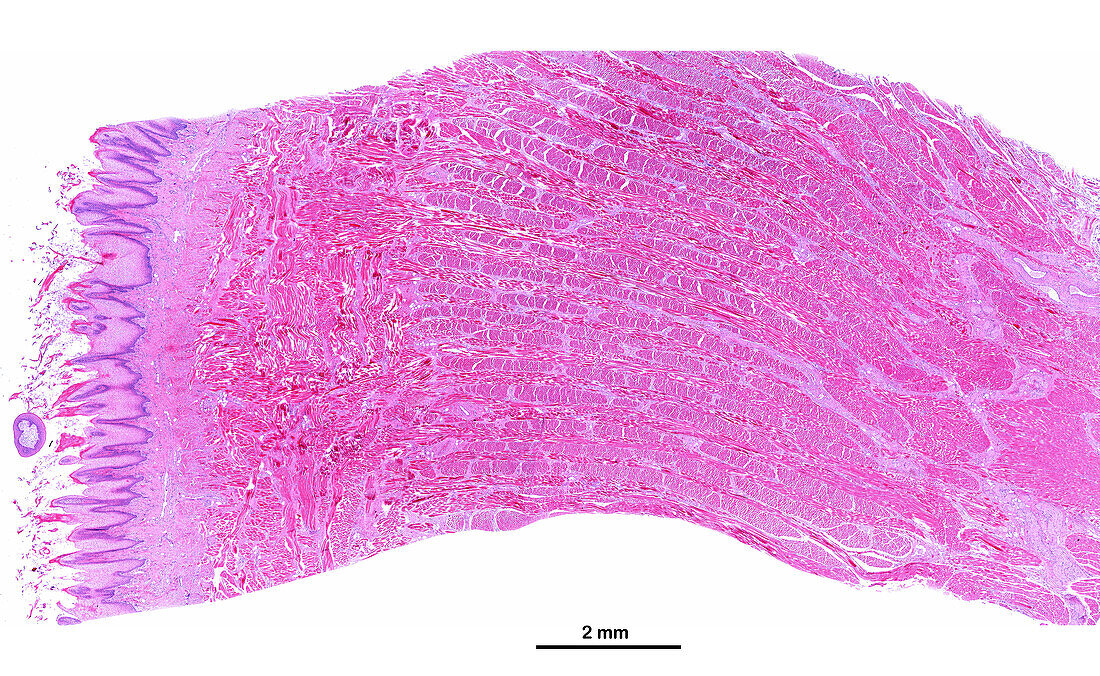
| Light micrograph of the upper surface of a human tongue showing, from left to right, the mucosa layer with many filiform papillae, the most numerous of the lingual papillae, a connective tissue lamina propria, and the muscle layer, with many intertwined striated muscle fibres. Haematoxylin and eosin stain. The scale bar corresponds to 2 mm. | |
| Licence : | Droits gérés |
| Crédit: | Science Photo Library / JOSE CALVO |
| Taille de l’image : | 4556 px × 2800 px |
| Model Release : | Non requis |
| Property Release : | Non requis |
| Restrictions : | - |
Prix pour cette image À partir de 45 €
Produit vendu
(Calendrier, Carte postale, Carte de vœux, Impression sur textile, Packaging etc)
À partir de 45 €
Usage commercial
(Affichage, Annonce presse, Annonce TV, Carte, Digital - hors rés. sociaux, Digital - rés. sociaux etc)
À partir de 45 €
Éditorial
(Digital, Journal, Livre, Livre pratique, Magazine, Télévision etc)
À partir de 60 €
Usage non-commercial
(Digital - hors rés. sociaux, Digital - rés. sociaux etc)
À partir de 120 €