Kidney, light micrograph
Numéro d’image : 13633585
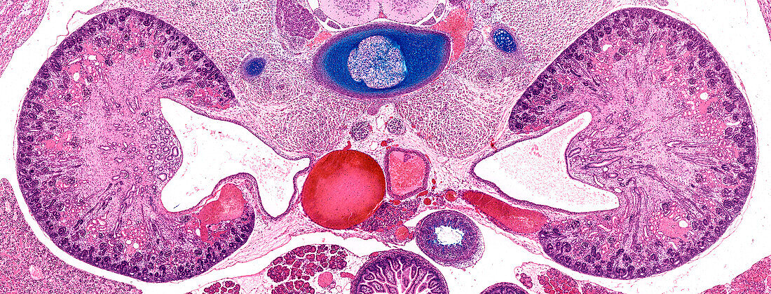
| Light micrograph of the developing kidneys of a foetus showing primitive renal corpuscles located around the outer cortex. Developing renal tubules and collecting ducts converge medially towards the large empty space termed the renal pelvis. In the postnatal kidney urine passes into the ureter via the pelvis. The aorta and inferior vena cava are seen in the midline, and a developing cartilaginous vertebral body and nucleus pulposus are stained blue. Paraffin section, alcian blue, and haematoxylin and eosin stain. Magnification: x16 when width printed at 10cm. | |
| Licence : | Droits gérés |
| Crédit: | Science Photo Library / Microscape |
| Taille de l’image : | 7265 px × 2766 px |
| Model Release : | Non requis |
| Property Release : | Non requis |
| Restrictions : | - |
Prix pour cette image À partir de 45 €
Produit vendu
(Calendrier, Carte postale, Carte de vœux, Impression sur textile, Packaging etc)
À partir de 45 €
Usage commercial
(Affichage, Annonce presse, Annonce TV, Carte, Digital - hors rés. sociaux, Digital - rés. sociaux etc)
À partir de 45 €
Éditorial
(Digital, Journal, Livre, Livre pratique, Magazine, Télévision etc)
À partir de 60 €
Usage non-commercial
(Digital - hors rés. sociaux, Digital - rés. sociaux etc)
À partir de 120 €
Mots clés
- aucun,
- bassinet du rein,
- biologie,
- biologique,
- cavité pyélique,
- corps humain,
- corps vertébral,
- corpuscule rénal,
- en bonne santé,
- histologie,
- histologique,
- micrographie optique,
- microscope optique,
- microscope photonique,
- microscopie optique,
- microscopie photonique,
- normal,
- noyau gélatineux,
- noyau pulpeux,
- nucleus pulposus,
- pelvis rénal,
- personne,
- petit bassin du rein,
- pyélon,
- rein,
- rénale,
- sain