Synovial joint, light micrograph
Numéro d’image : 13633583
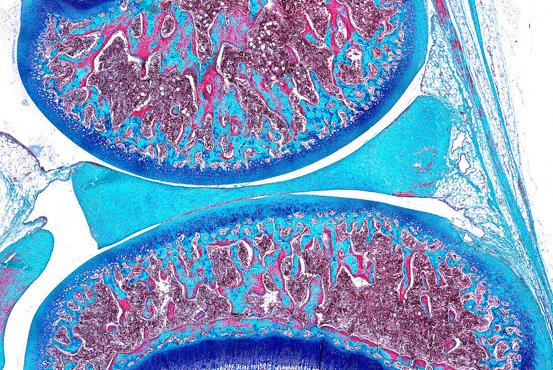
| Synovial joint, light micrograph. The joint space between articular bones of a synovial joint shows the meniscus composed of fibrocartilage. The bone ends show a surface of articular hyaline cartilage stained blue, deep to which is a wide region of developing bone (green) and bone marrow tissues (red). Paraffin section, trichrome stain. Magnification: x2 when width printed at 10cm. | |
| Licence : | Droits gérés |
| Crédit: | Science Photo Library / Microscape |
| Taille de l’image : | 6170 px × 4134 px |
| Model Release : | Non requis |
| Property Release : | Non requis |
| Restrictions : | - |
Prix pour cette image À partir de 45 €
Produit vendu
(Calendrier, Carte postale, Carte de vœux, Impression sur textile, Packaging etc)
À partir de 45 €
Usage commercial
(Affichage, Annonce presse, Annonce TV, Carte, Digital - hors rés. sociaux, Digital - rés. sociaux etc)
À partir de 45 €
Éditorial
(Digital, Journal, Livre, Livre pratique, Magazine, Télévision etc)
À partir de 60 €
Usage non-commercial
(Digital - hors rés. sociaux, Digital - rés. sociaux etc)
À partir de 120 €
Mots clés
- articulation synoviale,
- aucun,
- biologie,
- biologique,
- cartilage,
- cartilage articulaire,
- corps humain,
- en bonne santé,
- espace commun,
- fibrocartilage,
- histologie,
- histologique,
- ménisque,
- micrographie optique,
- microscope optique,
- microscope photonique,
- microscopie optique,
- microscopie photonique,
- normal,
- personne,
- sain