Seminiferous tubule, light micrograph
Numéro d’image : 13633581
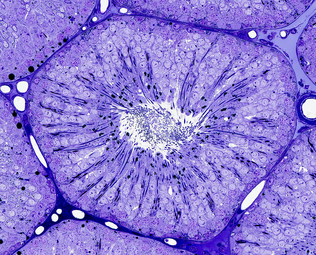
| Light micrograph of a seminiferous tubule in cross-section containing the seminiferous epithelium showing the process of spermatogenesis. Elongating spermatids form radially arranged columns between which are the earlier generations of maturing and proliferating germ cells. The tubule is supported by the surrounding intertubular tissue. Epoxy resin section, toluidine blue stain. Magnification: x40 when width printed at 10cm. | |
| Licence : | Droits gérés |
| Crédit: | Science Photo Library / Microscape |
| Taille de l’image : | 4697 px × 3780 px |
| Model Release : | Non requis |
| Property Release : | Non requis |
| Restrictions : | - |
Prix pour cette image À partir de 45 €
Produit vendu
(Calendrier, Carte postale, Carte de vœux, Impression sur textile, Packaging etc)
À partir de 45 €
Usage commercial
(Affichage, Annonce presse, Annonce TV, Carte, Digital - hors rés. sociaux, Digital - rés. sociaux etc)
À partir de 45 €
Éditorial
(Digital, Journal, Livre, Livre pratique, Magazine, Télévision etc)
À partir de 60 €
Usage non-commercial
(Digital - hors rés. sociaux, Digital - rés. sociaux etc)
À partir de 120 €
Mots clés
- aucun,
- biologie,
- biologique,
- cellules germinales,
- corps humain,
- en bonne santé,
- épithélium séminifère,
- germen,
- histologie,
- histologique,
- micrographie optique,
- microscope optique,
- microscope photonique,
- microscopie optique,
- microscopie photonique,
- normal,
- personne,
- sain,
- spermatogenèse,
- système reproducteur masculin,
- testicules,
- testis,
- tube séminifère