Human sperm, TEM.
Numéro d’image : 13620788
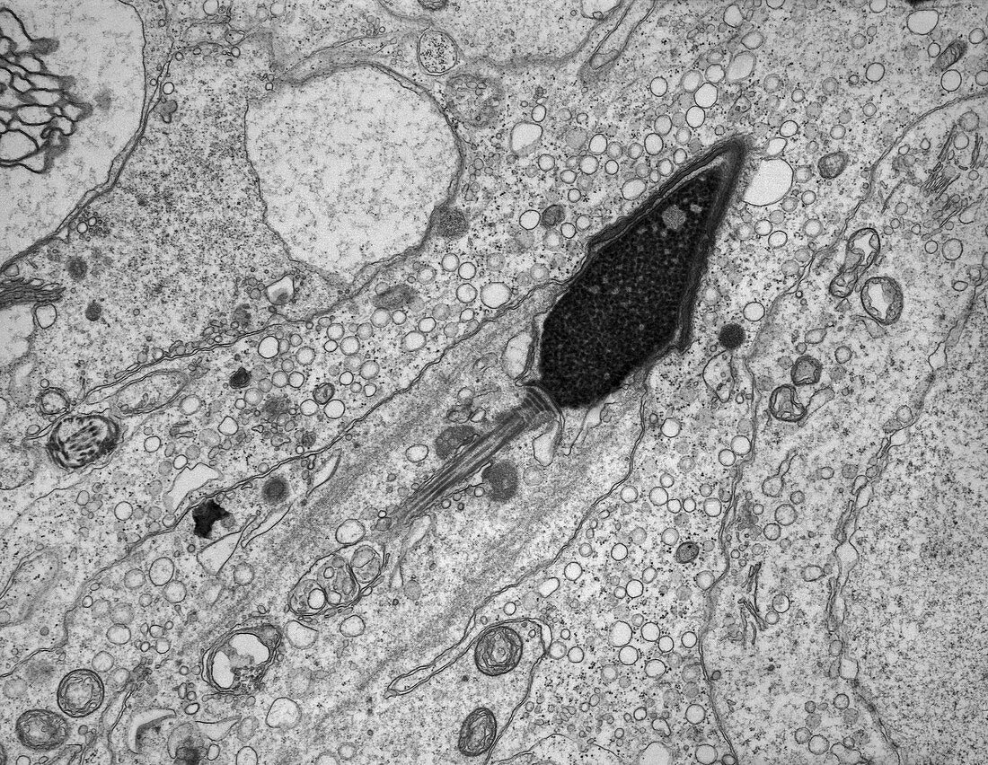
| Transmission electron micrograph (TEM) of the ultrastructure of an elongating human spermatid located within a column of Sertoli cell cytoplasm in the seminiferous epithelium. The cell DNA (deoxyribonucleic acid) has compacted into a pyriform-shaped nucleus to which is attached part of the developing sperm tail or flagellum. Covering the leading surface of the nucleus is the acrosome. The spermatid cytoplasm is being extruded distally and will ultimately be discarded when the sperm is released from the Sertoli cell in the process termed spermiation. Magnification: x8, 000 when height printed at 10cm. | |
| Licence : | Droits gérés |
| Crédit: | Science Photo Library / Microscape |
| Taille de l’image : | 4843 px × 3749 px |
| Model Release : | Non requis |
| Property Release : | Non requis |
| Restrictions : | - |
Prix pour cette image À partir de 45 €
Produit vendu
(Calendrier, Carte postale, Carte de vœux, Impression sur textile, Packaging etc)
À partir de 45 €
Usage commercial
(Affichage, Annonce presse, Annonce TV, Carte, Digital - hors rés. sociaux, Digital - rés. sociaux etc)
À partir de 45 €
Éditorial
(Digital, Journal, Livre, Livre pratique, Magazine, Télévision etc)
À partir de 60 €
Usage non-commercial
(Digital - hors rés. sociaux, Digital - rés. sociaux etc)
À partir de 120 €
Mots clés
- acrosome,
- aucun,
- biologie cellulaire,
- biologique,
- cytologie,
- cytologique,
- flagelle,
- flagellum,
- M.E.T.,
- MET,
- micrographie électronique,
- micrographie électronique à transmission,
- microscope électronique,
- microscope électronique à transmission,
- microscopie électronique,
- monochrome,
- n/b,
- noir et blanc,
- personne,
- spermatide,
- spermatogenèse,
- spermatozoïde,
- sperme humain,
- spermiogenèse,
- testicules,
- testis,
- ultrastructure,
- ultrastructure des cellules