Spermatogenesis in human testicle, light micrographs
Numéro d’image : 13613989
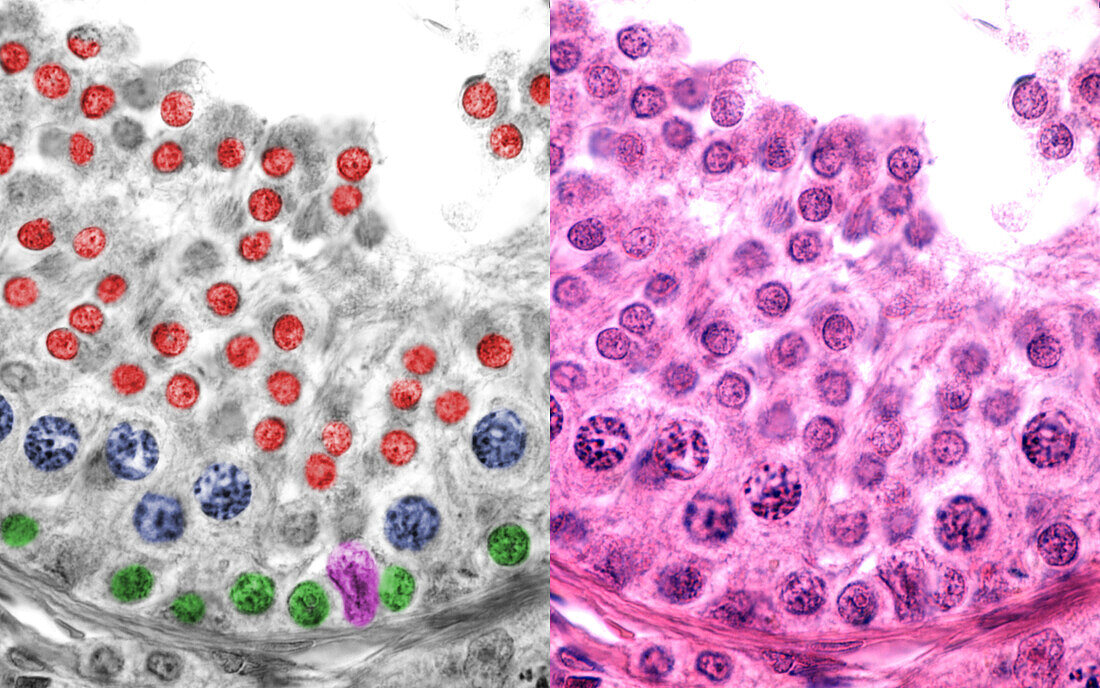
| Human testicle, light micrographs. The bottom micrograph shows a seminiferous tubule. In the top micrograph the cell types of the male germinal epithelium have been marked with colour. Seen are; Sertoli cells (pink), spermatogonia (green), primary spermatocytes in pachytene phase (blue) and spermatids (red). | |
| Licence : | Droits gérés |
| Crédit: | Science Photo Library / JOSE CALVO |
| Taille de l’image : | 6144 px × 3840 px |
| Model Release : | Non requis |
| Property Release : | Non requis |
| Restrictions : | - |
Prix pour cette image À partir de 45 €
Produit vendu
(Calendrier, Carte postale, Carte de vœux, Impression sur textile, Packaging etc)
À partir de 45 €
Usage commercial
(Affichage, Annonce presse, Annonce TV, Carte, Digital - hors rés. sociaux, Digital - rés. sociaux etc)
À partir de 45 €
Éditorial
(Digital, Journal, Livre, Livre pratique, Magazine, Télévision etc)
À partir de 60 €
Usage non-commercial
(Digital - hors rés. sociaux, Digital - rés. sociaux etc)
À partir de 120 €
Mots clés
- aucun,
- biologie,
- biologique,
- coloré,
- colorié,
- colorisé,
- corps humain,
- en bonne santé,
- histologie,
- histologique,
- humain,
- Leydig,
- méiose,
- micrographe,
- micrographie optique,
- microscope,
- microscope optique,
- microscopie,
- microscopie optique,
- normal,
- personne,
- sain,
- séminifère,
- spermatide,
- spermatocyte,
- spermatogenèse,
- spermatogonie,
- spermatozoa,
- spermatozoïdes,
- testicule