Human testis, TEM
Numéro d’image : 13613429
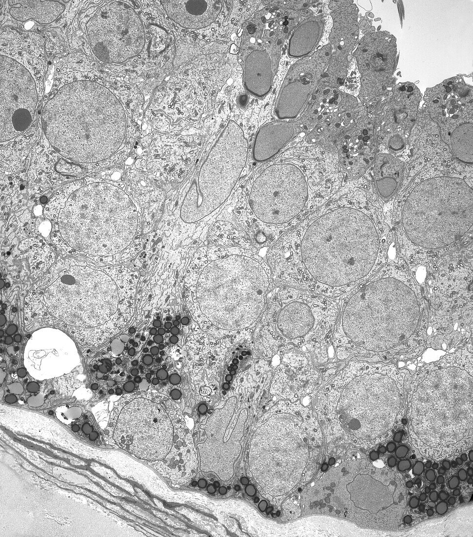
| Transmission electron micrograph (TEM) of the ultrastructure of the seminiferous epithelium of the human testis. Near the basal lamina is a Sertoli cell with many lipid inclusions (grey circles) and a spermatogonium and a primary spermatocyte. Another Sertoli cell nucleus is seen within a column of its own cytoplasm. At this higher level are numerous large rounded primary spermatocytes which are supported by ramifications of Sertoli cell cytoplasm. More apically are elongating spermatids with acrosomal caps covering the cranial aspect of their nuclei. Magnification: x1, 000 when height printed at 10cm. | |
| Licence : | Droits gérés |
| Crédit: | Science Photo Library / Microscape |
| Taille de l’image : | 3898 px × 4421 px |
| Model Release : | Non requis |
| Property Release : | Non requis |
| Restrictions : | - |
Prix pour cette image À partir de 45 €
Produit vendu
(Calendrier, Carte postale, Carte de vœux, Impression sur textile, Packaging etc)
À partir de 45 €
Usage commercial
(Affichage, Annonce presse, Annonce TV, Carte, Digital - hors rés. sociaux, Digital - rés. sociaux etc)
À partir de 45 €
Éditorial
(Digital, Journal, Livre, Livre pratique, Magazine, Télévision etc)
À partir de 60 €
Usage non-commercial
(Digital - hors rés. sociaux, Digital - rés. sociaux etc)
À partir de 120 €
Mots clés
- acrosome,
- aucun,
- biologie cellulaire,
- biologique,
- cellule de Sertoli,
- cytologie,
- cytologique,
- épithélium séminifère,
- M.E.T.,
- masculin,
- MET,
- micrographie électronique,
- micrographie électronique à transmission,
- microscope électronique,
- microscope électronique à transmission,
- microscopie électronique,
- monochrome,
- n/b,
- noir et blanc,
- personne,
- spermatide,
- spermatocyte,
- spermatogenèse,
- testicules,
- testis,
- ultrastructure