Kidney glomerulus, light micrograph
Numéro d’image : 13586057
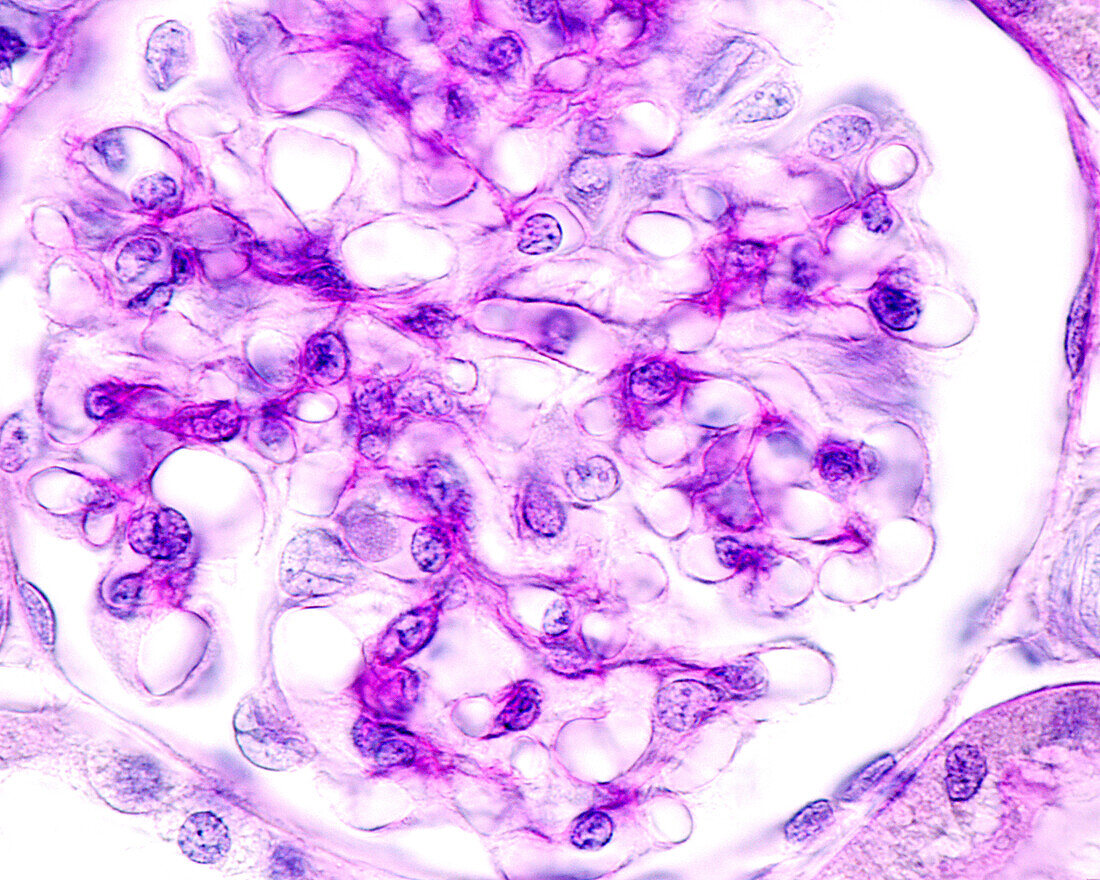
| Light micrograph of a kidney glomerulus stained with the periodic acid Schiff (PAS) method. The vascular tuft, located in the centre of the glomerulus, shows intense staining with PAS, which corresponds to the basement membranes of the glomerular capillaries. In many kidney diseases, the initial modifications consist of changes in this basement membrane of the glomerular capillaries, which can be detected using the PAS method. | |
| Licence : | Droits gérés |
| Crédit: | Science Photo Library / JOSE CALVO |
| Taille de l’image : | 3840 px × 3072 px |
| Model Release : | Non requis |
| Property Release : | Non requis |
| Restrictions : | - |
Prix pour cette image À partir de 45 €
Produit vendu
(Calendrier, Carte postale, Carte de vœux, Impression sur textile, Packaging etc)
À partir de 45 €
Usage commercial
(Affichage, Annonce presse, Annonce TV, Carte, Digital - hors rés. sociaux, Digital - rés. sociaux etc)
À partir de 45 €
Éditorial
(Digital, Journal, Livre, Livre pratique, Magazine, Télévision etc)
À partir de 60 €
Usage non-commercial
(Digital - hors rés. sociaux, Digital - rés. sociaux etc)
À partir de 120 €