Kidney cortex, light micrograph
Numéro d’image : 13586046
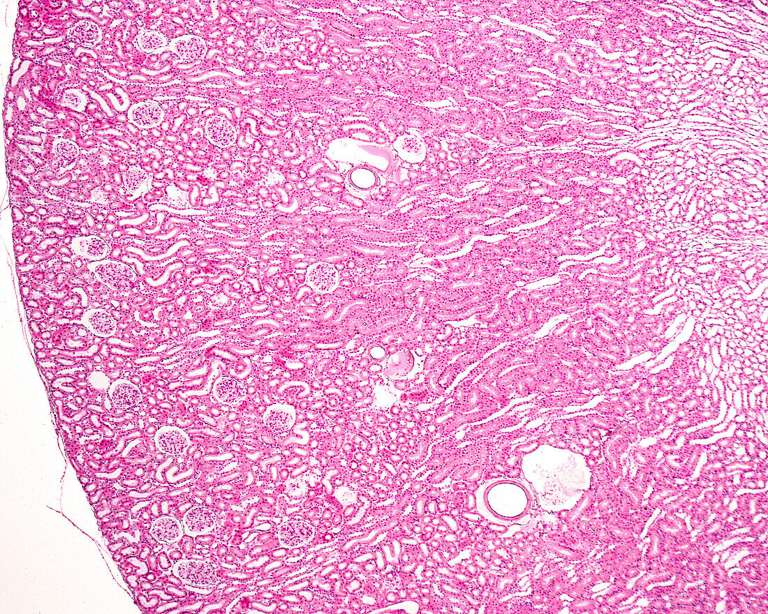
| Light micrograph of the renal cortex of an albino rat. The smaller size of the kidney makes it possible to observe the entire renal cortex and the border between the cortex and medulla in a single microscopic field. The tissue was fixed by perfusion, which explains the dilated appearance of the cortical arteries and veins. In the cortex, renal corpuscles are clearly visible. In the outer medulla (right), the difference between the outer stripe (similar to the cortex, but without renal corpuscles) and the inner stripe (which appears lighter, with cross-sectioned tubules) can be seen. | |
| Licence : | Droits gérés |
| Crédit: | Science Photo Library / JOSE CALVO |
| Taille de l’image : | 3840 px × 3072 px |
| Model Release : | Non requis |
| Property Release : | Non requis |
| Restrictions : | - |
Prix pour cette image À partir de 45 €
Produit vendu
(Calendrier, Carte postale, Carte de vœux, Impression sur textile, Packaging etc)
À partir de 45 €
Usage commercial
(Affichage, Annonce presse, Annonce TV, Carte, Digital - hors rés. sociaux, Digital - rés. sociaux etc)
À partir de 45 €
Éditorial
(Digital, Journal, Livre, Livre pratique, Magazine, Télévision etc)
À partir de 60 €
Usage non-commercial
(Digital - hors rés. sociaux, Digital - rés. sociaux etc)
À partir de 120 €
Mots clés
- anatomie,
- anatomique,
- animal,
- aucun,
- biologie,
- cortex rénal,
- glomérule,
- glomerulus,
- histologie,
- histologique,
- médullaire rénale,
- micrographie,
- microscope,
- microscope optique,
- microscopie,
- microscopie optique,
- microscopique,
- néphron,
- personne,
- rayons médullaires,
- rein,
- renal medulla,
- rénale,
- tubule complexe,
- tubule tordu,
- urinaire