Human pineal gland lobulation, light micrograph
Numéro d’image : 13585792
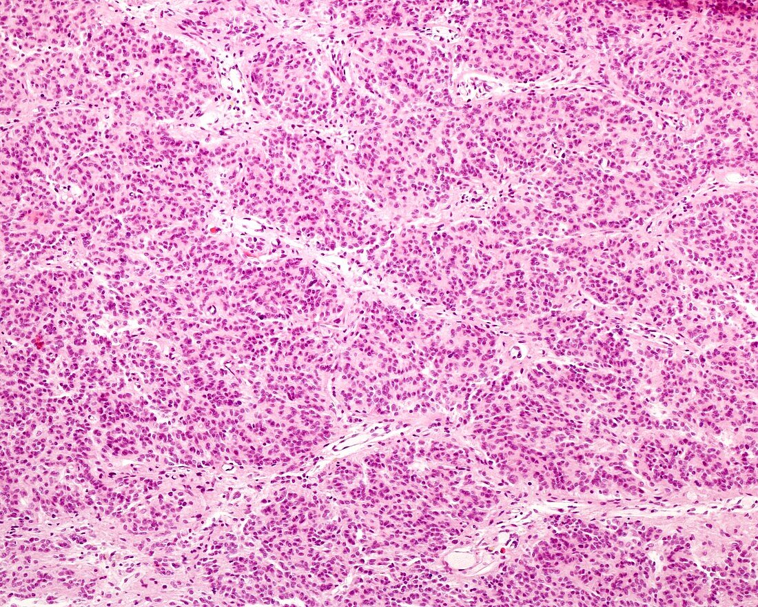
| Light micrograph of a young human pineal gland showing incipient lobulation of the parenchyma. The pineal parenchyma tends to organize itself into rounded units separated by thin connective tissue septa where the blood vessels are located. However, unlike the true lobes present in the exocrine glands, these units are not delimited but rather merge extensively with neighbouring units. Although the distribution of the parenchyma in rounded units can be seen, the lobes are still poorly defined. | |
| Licence : | Droits gérés |
| Crédit: | Science Photo Library / JOSE CALVO |
| Taille de l’image : | 3840 px × 3072 px |
| Model Release : | Non requis |
| Property Release : | Non requis |
| Restrictions : | - |
Prix pour cette image À partir de 45 €
Produit vendu
(Calendrier, Carte postale, Carte de vœux, Impression sur textile, Packaging etc)
À partir de 45 €
Usage commercial
(Affichage, Annonce presse, Annonce TV, Carte, Digital - hors rés. sociaux, Digital - rés. sociaux etc)
À partir de 45 €
Éditorial
(Digital, Journal, Livre, Livre pratique, Magazine, Télévision etc)
À partir de 60 €
Usage non-commercial
(Digital - hors rés. sociaux, Digital - rés. sociaux etc)
À partir de 120 €