Human adrenal gland cortex and medulla, light micrograph
Numéro d’image : 13585232
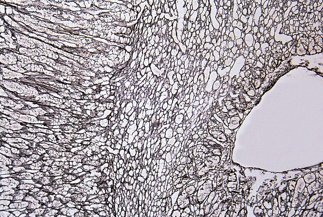
| Light micrograph of an adrenal gland stained with a silver method to demonstrate reticular fibres. The cortex occupies the left third of the image. The rest is adrenal medulla. The different thickness and orientation of the cellular cords between the cortex and the medulla clearly stand out. The empty space at far right is a vein. | |
| Licence : | Droits gérés |
| Crédit: | Science Photo Library / JOSE CALVO |
| Taille de l’image : | 3840 px × 2575 px |
| Model Release : | Non requis |
| Property Release : | Non requis |
| Restrictions : | - |
Prix pour cette image À partir de 45 €
Produit vendu
(Calendrier, Carte postale, Carte de vœux, Impression sur textile, Packaging etc)
À partir de 45 €
Usage commercial
(Affichage, Annonce presse, Annonce TV, Carte, Digital - hors rés. sociaux, Digital - rés. sociaux etc)
À partir de 45 €
Éditorial
(Digital, Journal, Livre, Livre pratique, Magazine, Télévision etc)
À partir de 60 €
Usage non-commercial
(Digital - hors rés. sociaux, Digital - rés. sociaux etc)
À partir de 120 €
Mots clés
- argent,
- aucun,
- biologie,
- biologique,
- cellule,
- cortex surrénal,
- cortico-surrénale,
- en bonne santé,
- endocrine,
- glande,
- histologie,
- histologique,
- medulla,
- médullaire,
- micrographie optique,
- microscope,
- microscope optique,
- microscopie,
- microscopie optique,
- microscopique,
- mitose,
- normal,
- personne,
- sain,
- surrénal,
- tissus