Poliovirus Type 1 Mahoney capsid, molecular model
Numéro d’image : 13505228
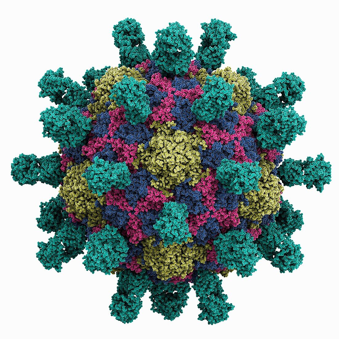
| Poliovirus Type 1 Mahoney capsid, molecular model. The image shows the capsid proteins VP1 (yellow), VP2 (pink), VP3 (blue) and the anti-VP1 monoclonal antibody (cyan). The VP4 protein is buried inside the capsid. | |
| Licence : | Droits gérés |
| Crédit: | Science Photo Library / Laguna Design |
| Taille de l’image : | 4150 px × 4150 px |
| Model Release : | Non requis |
| Property Release : | Non requis |
| Restrictions : | - |
Prix pour cette image À partir de 45 €
Produit vendu
(Calendrier, Carte postale, Carte de vœux, Impression sur textile, Packaging etc)
À partir de 45 €
Usage commercial
(Affichage, Annonce presse, Annonce TV, Carte, Digital - hors rés. sociaux, Digital - rés. sociaux etc)
À partir de 45 €
Éditorial
(Digital, Journal, Livre, Livre pratique, Magazine, Télévision etc)
À partir de 60 €
Usage non-commercial
(Digital - hors rés. sociaux, Digital - rés. sociaux etc)
À partir de 120 €
Mots clés
- anticorps,
- arrière plan blanc,
- arrière-plan blanc,
- aucun,
- biochimie,
- biochimique,
- biologie,
- biologique,
- capside,
- espace rempli,
- fond blanc,
- illustration,
- modèle moléculaire,
- moléculaire,
- molécule,
- monoclonal,
- oeuvre,
- personne,
- polio,
- protéine,
- remplissage d'espace,
- structure,
- structure moléculaire,
- type 1,
- virus,
- virus de la polio