Trachea, LM
Numéro d’image : 13496809
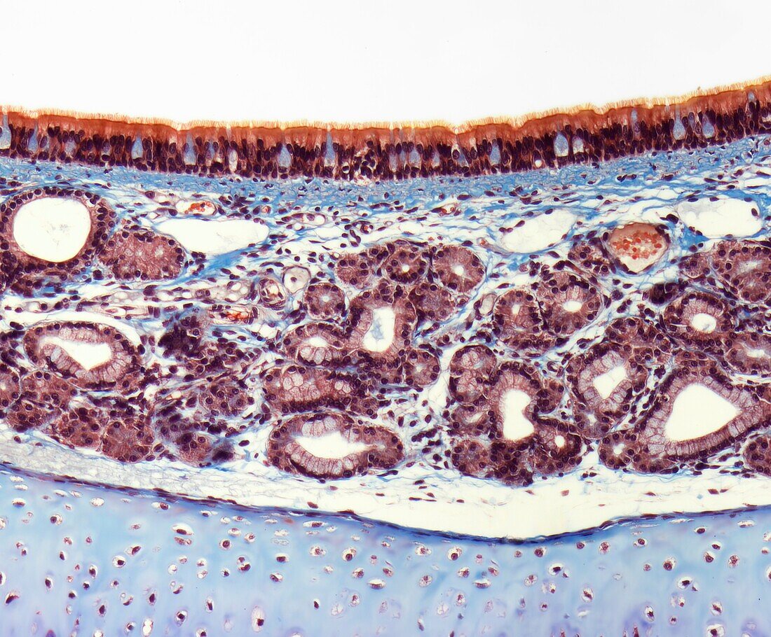
| Trachea. Light micrograph (LM) of a transverse section through the trachea. At the top are the columnar epithelial cells that line the trachea. They have hair-like cilia on their surface that beat to move mucus, and particles trapped in it, upwards out of the respiratory tract. The mucus produced by mucosa glands (red, hollow) and secreted by goblet cells in the epithelium.Magnification: 150 when printed at 10 centimetres wide. | |
| Licence : | Droits gérés |
| Crédit: | Science Photo Library / Gschmeissner, Steve |
| Taille de l’image : | 4596 px × 3802 px |
| Model Release : | Non requis |
| Property Release : | Non requis |
| Restrictions : | - |
Prix pour cette image À partir de 45 €
Produit vendu
(Calendrier, Carte postale, Carte de vœux, Impression sur textile, Packaging etc)
À partir de 45 €
Usage commercial
(Affichage, Annonce presse, Annonce TV, Carte, Digital - hors rés. sociaux, Digital - rés. sociaux etc)
À partir de 45 €
Éditorial
(Digital, Journal, Livre, Livre pratique, Magazine, Télévision etc)
À partir de 60 €
Usage non-commercial
(Digital - hors rés. sociaux, Digital - rés. sociaux etc)
À partir de 120 €
Mots clés
- anatomie,
- anatomique,
- biologie,
- biologique,
- catégorie,
- cellule à mucus,
- cellule caliciforme,
- cellule en gobelet,
- cellule mucipare,
- cellule muqueuse à pôle apical ouvert,
- cils,
- corps humain,
- coupe,
- épithélium cylindrique,
- histologie,
- histologique,
- humain,
- médecine,
- médical,
- médicale,
- micrographie optique,
- microscope optique,
- microscopie optique,
- mucus,
- muqueuse,
- muqueux,
- partie,
- respiratoire,
- section,
- tissu humain,
- trachea,
- trachée