Bladder, LM
Numéro d’image : 13496785
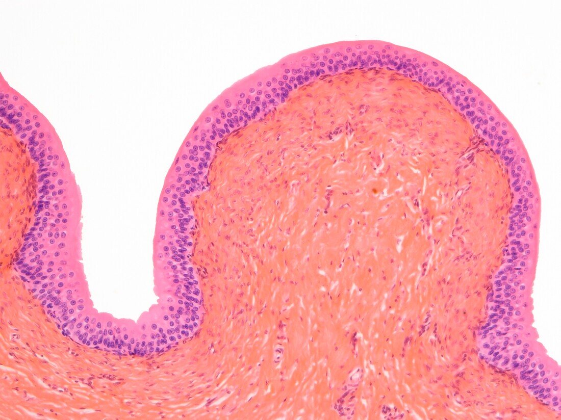
| Bladder. Light micrograph (LM) of a vertical section through the wall of the urinary bladder. The inner surface is at top. The upper layer (magenta) is the transitional epithelium, which is especially adapted for the bladder's function of containing urine. The plasma membranes surrounding the epithelial cells here are much thicker than most cell membranes, with a specialised sub-structure. The epithelium is thus rendered impermeable to potentially toxic urine. Below the epithelium is smooth muscle and connective tissue (pink). Magnification: x150 when printed 10 centimetres wide. | |
| Licence : | Droits gérés |
| Crédit: | Science Photo Library / Gschmeissner, Steve |
| Taille de l’image : | 4829 px × 3619 px |
| Model Release : | Non requis |
| Restrictions : | - |
Prix pour cette image À partir de 45 €
Produit vendu
(Calendrier, Carte postale, Carte de vœux, Impression sur textile, Packaging etc)
À partir de 45 €
Usage commercial
(Affichage, Annonce presse, Annonce TV, Carte, Digital - hors rés. sociaux, Digital - rés. sociaux etc)
À partir de 45 €
Éditorial
(Digital, Journal, Livre, Livre pratique, Magazine, Télévision etc)
À partir de 60 €
Usage non-commercial
(Digital - hors rés. sociaux, Digital - rés. sociaux etc)
À partir de 120 €