Pleomorphic Xanthoastrocytoma MR
Numéro d’image : 13496226
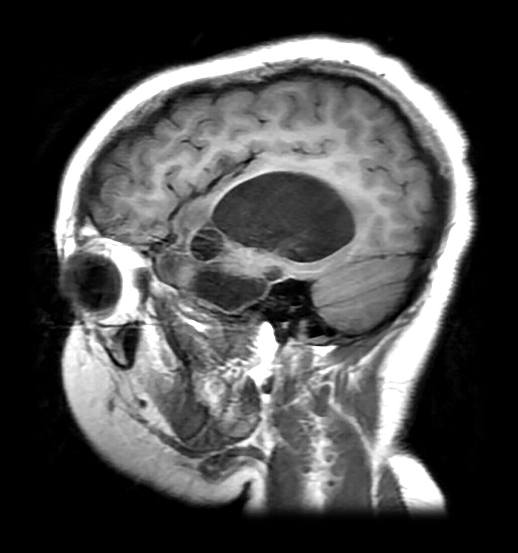
| This sagittal (from the side) T1 weighted MR image without contrast shows a multicystic tumour in the temporal lobe with mass effect which represents a low grade tumour called a pleomorphic xanthoastrocytoma. These are often located in the temporal lobe in younger patients and associated with chronic seizures. | |
| Licence : | Droits gérés |
| Crédit: | Science Photo Library / Science Source / Living Art Enterprises, LLC |
| Taille de l’image : | 3900 px × 4161 px |
| Model Release : | Non requis |
| Property Release : | Non requis |
| Restrictions : | - |
Prix pour cette image À partir de 45 €
Produit vendu
(Calendrier, Carte postale, Carte de vœux, Impression sur textile, Packaging etc)
À partir de 45 €
Usage commercial
(Affichage, Annonce presse, Annonce TV, Carte, Digital - hors rés. sociaux, Digital - rés. sociaux etc)
À partir de 45 €
Éditorial
(Digital, Journal, Livre, Livre pratique, Magazine, Télévision etc)
À partir de 60 €
Usage non-commercial
(Digital - hors rés. sociaux, Digital - rés. sociaux etc)
À partir de 120 €