Aqueductal Stenosis, Hydrocephalus, MRI
Numéro d’image : 13496131
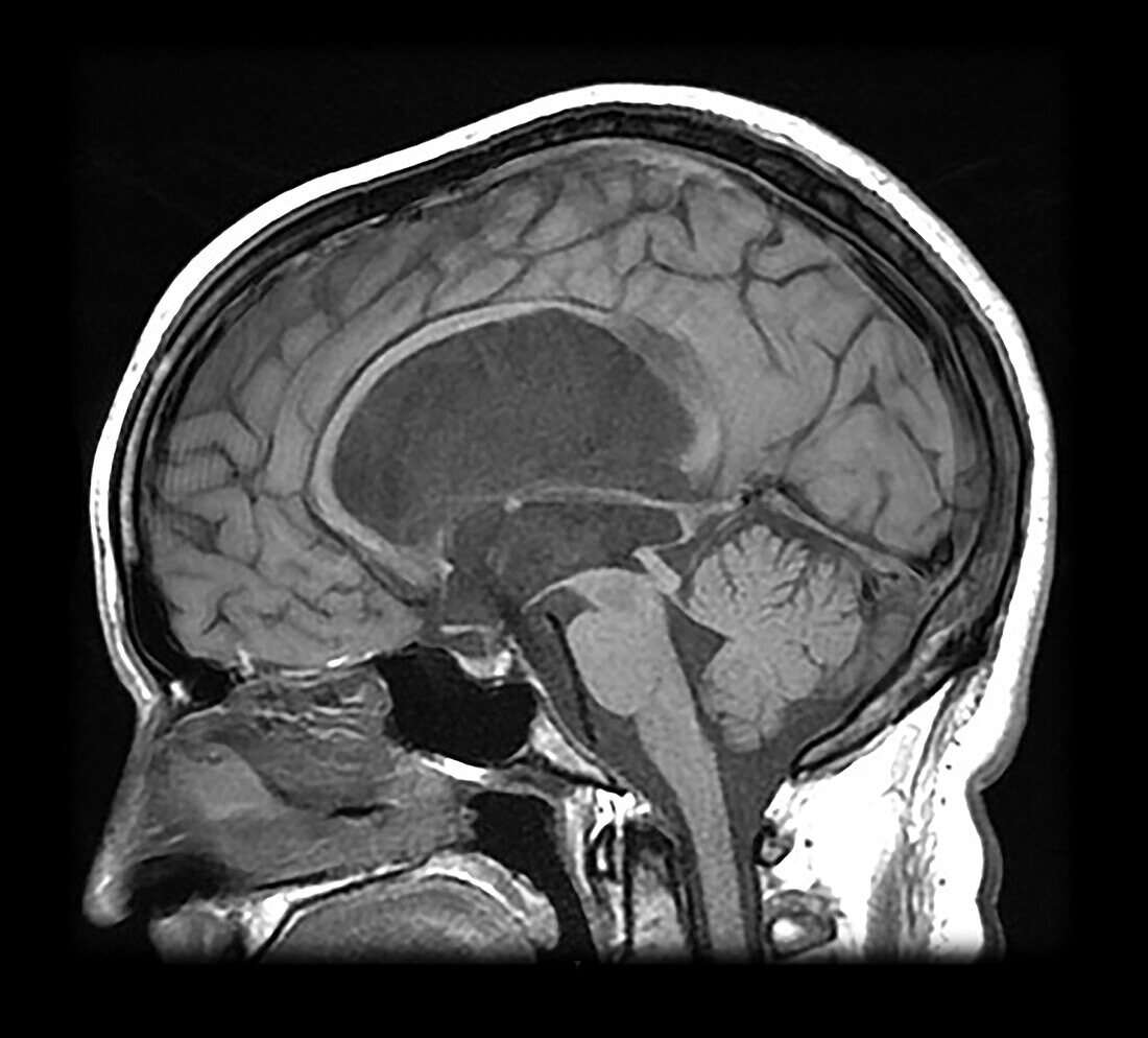
| This sagittal T1 weighted MR image shows dilated third and lateral ventricles with a normal size fourth ventricle. Stenosis with occlusion of the lower aqueduct of Sylvius is present. Surgical intervention has been performed with an inferior third ventriculostomy which is patent as demonstrated by a prominent flow void of CSF flow from the third ventricle to the pre-pontine cistern. | |
| Licence : | Droits gérés |
| Crédit: | Science Photo Library / Science Source / Living Art Enterprises, LLC |
| Taille de l’image : | 4318 px × 3900 px |
| Model Release : | Non requis |
| Property Release : | Non requis |
| Restrictions : | - |
Prix pour cette image À partir de 45 €
Produit vendu
(Calendrier, Carte postale, Carte de vœux, Impression sur textile, Packaging etc)
À partir de 45 €
Usage commercial
(Affichage, Annonce presse, Annonce TV, Carte, Digital - hors rés. sociaux, Digital - rés. sociaux etc)
À partir de 45 €
Éditorial
(Digital, Journal, Livre, Livre pratique, Magazine, Télévision etc)
À partir de 60 €
Usage non-commercial
(Digital - hors rés. sociaux, Digital - rés. sociaux etc)
À partir de 120 €