Extensive Chronic Allergic Fungal Sinusitis
Numéro d’image : 13496085
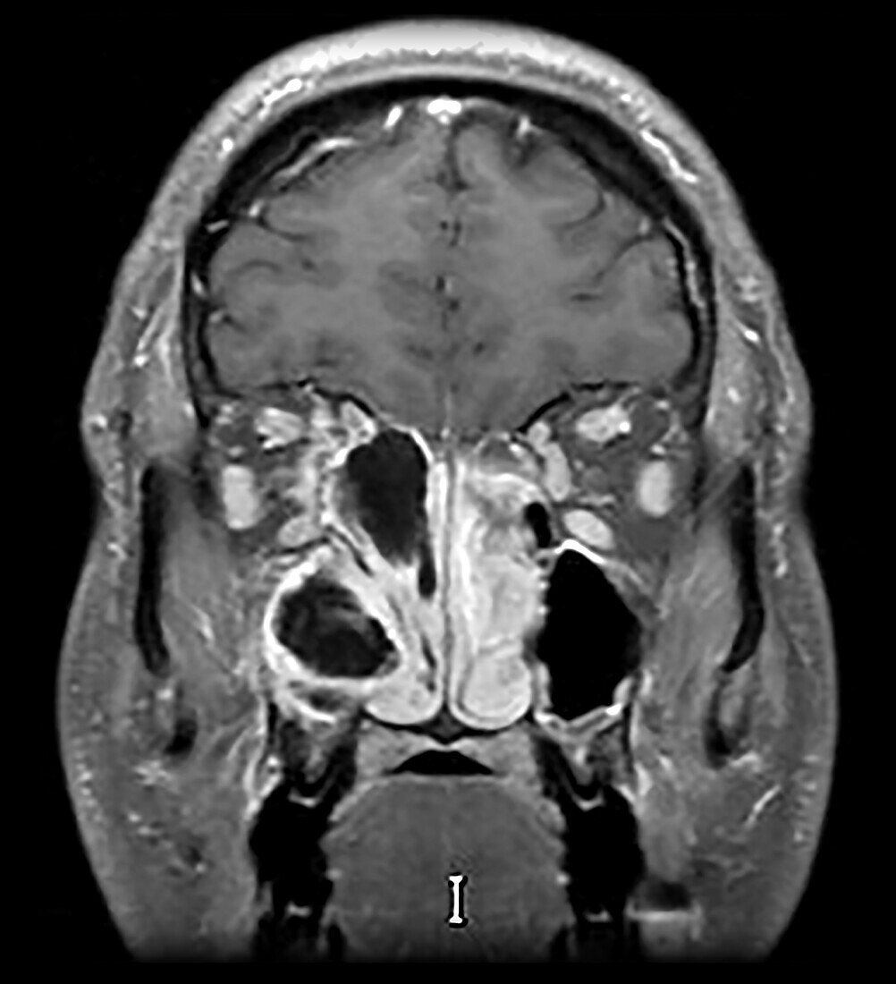
| This coronal (frontal view) contrast enhanced T1 weighted MR image shows what appears to be relatively aerated paranasal sinuses with mild mucosal thickening which in fact represents extensive fungal sinusitis with marked increase protein content within opacified sinuses. The elevated protein mimics aerated sinuses. The involved ethmoid air cells are expanded, extending into the orbit on the viewers left. | |
| Licence : | Droits gérés |
| Crédit: | Science Photo Library / Science Source / Living Art Enterprises, LLC |
| Taille de l’image : | 3900 px × 4280 px |
| Model Release : | Non requis |
| Property Release : | Non requis |
| Restrictions : | - |
Prix pour cette image À partir de 45 €
Produit vendu
(Calendrier, Carte postale, Carte de vœux, Impression sur textile, Packaging etc)
À partir de 45 €
Usage commercial
(Affichage, Annonce presse, Annonce TV, Carte, Digital - hors rés. sociaux, Digital - rés. sociaux etc)
À partir de 45 €
Éditorial
(Digital, Journal, Livre, Livre pratique, Magazine, Télévision etc)
À partir de 60 €
Usage non-commercial
(Digital - hors rés. sociaux, Digital - rés. sociaux etc)
À partir de 120 €