Pituitary gland, light micrograph
Numéro d’image : 13478078
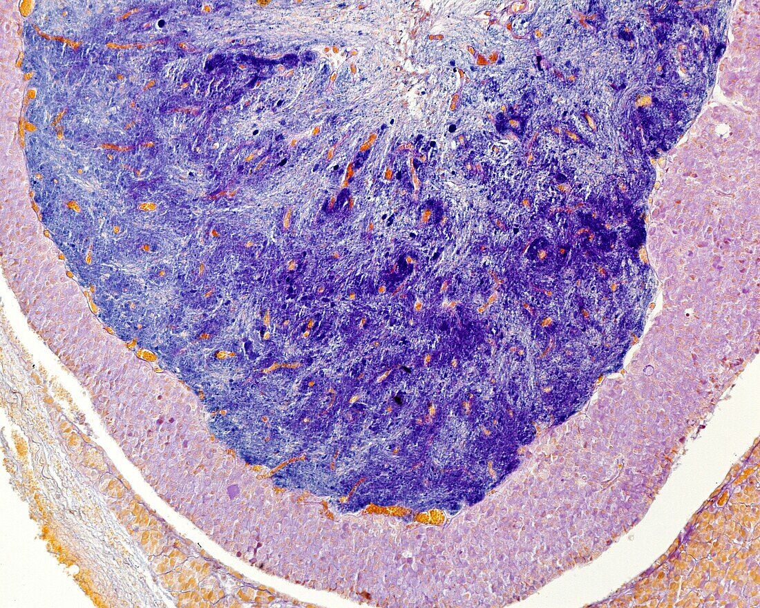
| Light micrograph of a cat pituitary gland showing the posterior lobe (top) stained with a method for neurosecretion. Below is the pars intermedia, Rathke's cleft (well visible in this species) and a small portion of the anterior pituitary. The neurosecretory material is distributed throughout the posterior pituitary showing a clear tendency to accumulate in the vicinity of the blood vessels. Some Herring bodies are observed. The orange-stained structures are blood vessels filled with red blood cells. Paraldehyde-thionine. | |
| Licence : | Droits gérés |
| Crédit: | Science Photo Library / JOSE CALVO |
| Taille de l’image : | 3840 px × 3072 px |
| Model Release : | Non requis |
| Property Release : | Non requis |
| Restrictions : | - |
Prix pour cette image À partir de 45 €
Produit vendu
(Calendrier, Carte postale, Carte de vœux, Impression sur textile, Packaging etc)
À partir de 45 €
Usage commercial
(Affichage, Annonce presse, Annonce TV, Carte, Digital - hors rés. sociaux, Digital - rés. sociaux etc)
À partir de 45 €
Éditorial
(Digital, Journal, Livre, Livre pratique, Magazine, Télévision etc)
À partir de 60 €
Usage non-commercial
(Digital - hors rés. sociaux, Digital - rés. sociaux etc)
À partir de 120 €
Mots clés
- anatomie,
- aucun,
- biologie,
- biologique,
- catégorie,
- corps humain,
- coupe,
- endocrinologie,
- glande,
- glande endocrinale,
- glande endocrine,
- glandulaire,
- histologie,
- histologique,
- hormone,
- hypophyse,
- hypophyse postérieure,
- hypothalamus,
- microscope,
- microscope optique,
- microscopie,
- microscopie optique,
- neurohypophyse,
- partie,
- personne,
- pituitaire arrière,
- pituitaire postérieur,
- posthypophyse,
- sécréteur,
- sécrétion,
- sécrétoire,
- section