Anatomy of the stem of Lamium purpureum
Numéro d’image : 13471202
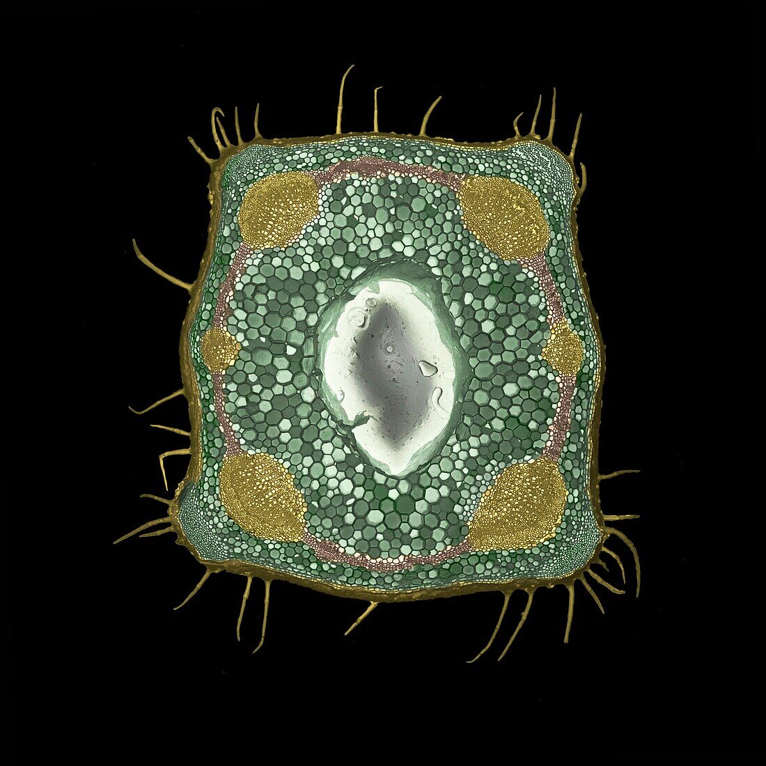
| Scanning electron micrograph of a stem of red dead nettle, Lamium purpureum in cross section, illustrating different tissue types. The picture shows the square outline of a hollow stem, with hairs on its surface. The coloured ring comprises six (yellow) water-conducting vascular bundles linked by a layer of cambium (brown). To the outside of the ring is the cortex; to the inside is the pith (both green). The pith consists of parenchyma - cells with no specialised function, that may contain chloroplasts or starch grains. During growth, the pith cells degenerate, giving rise to the hollow stem here. The hollow space is not empty; it is filled with water and remnants of cell walls. The cortex is also parenchymatous, but at the extreme corners, it has differentiated to form a tissue of small cells called collenchyma. This is a structural specialisation giving mechanical support to the stem | |
| Licence : | Droits gérés |
| Crédit: | Science Photo Library / Burgess, Dr. Jeremy |
| Taille de l’image : | 4500 px × 4500 px |
| Model Release : | Non requis |
| Restrictions : | - |
Prix pour cette image À partir de 45 €
Produit vendu
(Calendrier, Carte postale, Carte de vœux, Impression sur textile, Packaging etc)
À partir de 45 €
Usage commercial
(Affichage, Annonce presse, Annonce TV, Carte, Digital - hors rés. sociaux, Digital - rés. sociaux etc)
À partir de 45 €
Éditorial
(Digital, Journal, Livre, Livre pratique, Magazine, Télévision etc)
À partir de 60 €
Usage non-commercial
(Digital - hors rés. sociaux, Digital - rés. sociaux etc)
À partir de 120 €