T wave changes, illustration
Numéro d’image : 13453416
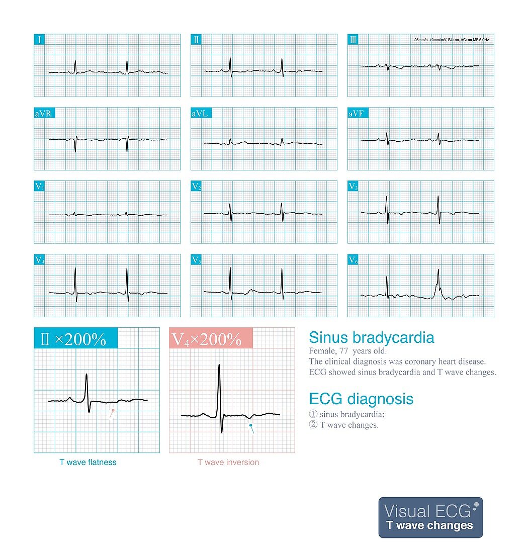
| Electrocardiogram (ECG) illustration showing T wave changes in a 77 year old woman diagnosed with coronary heart disease. ECG showed sinus bradycardia and T wave changes. The abnormal T waves are mainly flat and inverted. The ECG artifact in lead V6 should not be misdiagnosed as ventricular premature beat. | |
| Licence : | Droits gérés |
| Crédit: | Science Photo Library / CHONGQING TUMI TECHNOLOGY LTD |
| Taille de l’image : | 3600 px × 3772 px |
| Model Release : | Non requis |
| Property Release : | Non requis |
| Restrictions : | - |
Prix pour cette image À partir de 45 €
Produit vendu
(Calendrier, Carte postale, Carte de vœux, Impression sur textile, Packaging etc)
À partir de 45 €
Usage commercial
(Affichage, Annonce presse, Annonce TV, Carte, Digital - hors rés. sociaux, Digital - rés. sociaux etc)
À partir de 45 €
Éditorial
(Digital, Journal, Livre, Livre pratique, Magazine, Télévision etc)
À partir de 60 €
Usage non-commercial
(Digital - hors rés. sociaux, Digital - rés. sociaux etc)
À partir de 120 €
Mots clés
- anormal,
- arrhythmia,
- arythmie,
- aucun,
- cardiaque,
- cardiologie,
- cœur,
- créé digitalement,
- désordre,
- ECG,
- électrocardiogramme,
- électrophysiologie,
- état,
- illustration,
- maladie,
- maladies cardiovasculaires,
- malsain,
- médecine,
- médical,
- médicale,
- oeuvre,
- personne,
- produit digitalement,
- réalisé digitalement,
- soins de santé,
- trouble