Rheumatoid arthritis, X-ray
Numéro d’image : 13453110
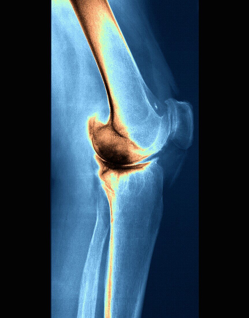
| X-ray image of the left knee of a 54-year-old female patient with rheumatoid arthritis. Rheumatoid arthritis is an autoimmune disease that attacks the joints, leading to painful, stiff and deformed joints. The scan shows diffuse bone demineralization producing a heterogeneous appearance by the existence of geodic images of internal femoral condylar and external and especially the internal tibial plateau. The patient also has bicompartmental knee osteoarthritis (osteoarthritis affecting two of three compartments of the knee joint). | |
| Licence : | Droits gérés |
| Crédit: | Science Photo Library / Zephyr |
| Taille de l’image : | 3402 px × 4336 px |
| Model Release : | Non requis |
| Property Release : | Non requis |
| Restrictions : | - |
Prix pour cette image À partir de 45 €
Produit vendu
(Calendrier, Carte postale, Carte de vœux, Impression sur textile, Packaging etc)
À partir de 45 €
Usage commercial
(Affichage, Annonce presse, Annonce TV, Carte, Digital - hors rés. sociaux, Digital - rés. sociaux etc)
À partir de 45 €
Éditorial
(Digital, Journal, Livre, Livre pratique, Magazine, Télévision etc)
À partir de 60 €
Usage non-commercial
(Digital - hors rés. sociaux, Digital - rés. sociaux etc)
À partir de 120 €