Stroke, MRI scan
Numéro d’image : 13452470
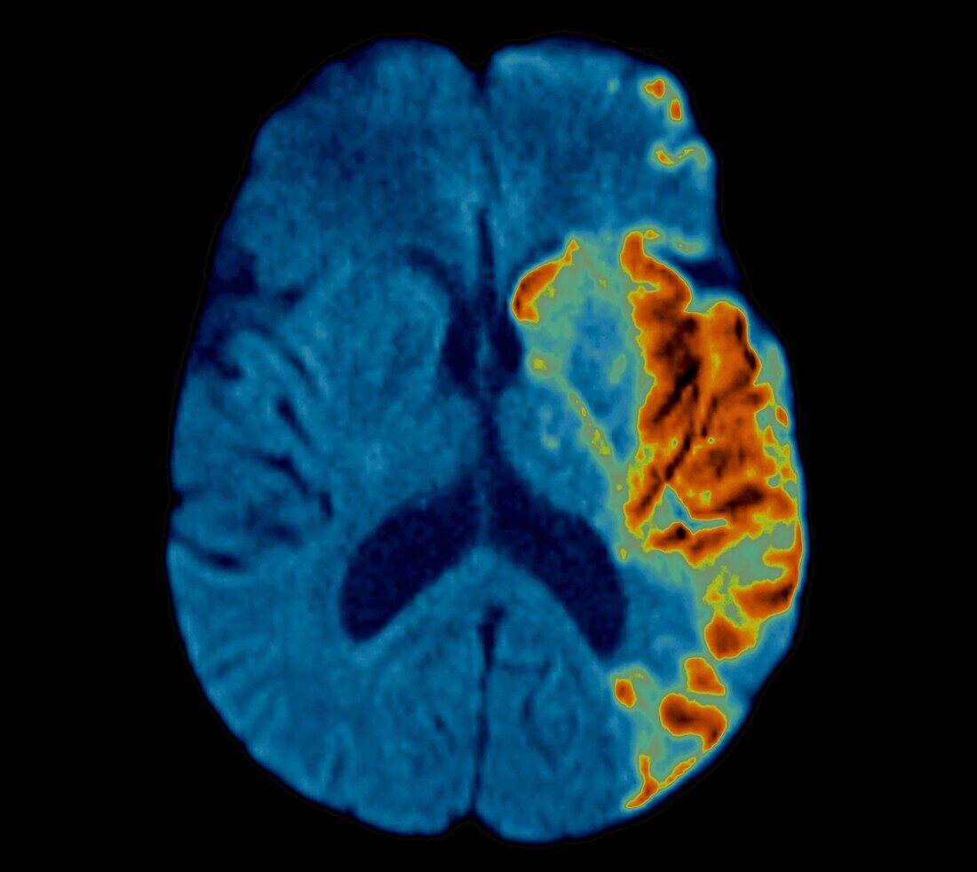
| Coloured axial magnetic resonance imaging (MRI) scan through the brain of a 66 year old male patient with a stroke. The damaged area of the brain is at lower right and centre right, on the left-hand side of the brain. There is a hypersignal (bright areas) of the caudate nucleus and cortex. This is a diffusion weighted imaging (DWI) MRI scan. | |
| Licence : | Droits gérés |
| Crédit: | Science Photo Library / Zephyr |
| Taille de l’image : | 3858 px × 3445 px |
| Model Release : | Non requis |
| Property Release : | Non requis |
| Restrictions : | - |
Prix pour cette image À partir de 45 €
Produit vendu
(Calendrier, Carte postale, Carte de vœux, Impression sur textile, Packaging etc)
À partir de 45 €
Usage commercial
(Affichage, Annonce presse, Annonce TV, Carte, Digital - hors rés. sociaux, Digital - rés. sociaux etc)
À partir de 45 €
Éditorial
(Digital, Journal, Livre, Livre pratique, Magazine, Télévision etc)
À partir de 60 €
Usage non-commercial
(Digital - hors rés. sociaux, Digital - rés. sociaux etc)
À partir de 120 €
Mots clés
- A.V.C,
- A.V.C.,
- accident cérébrovasculaire,
- accident vasculaire cérébral,
- anormal,
- arrière plan noir,
- arrière-plan noir,
- artère cérébrale moyenne,
- attaque cérébrale,
- aucun,
- AVC,
- axial,
- cortex,
- fond noir,
- homme,
- I.R.M.,
- imagerie par résonance magnétique,
- imagerie par résonnance magnétique,
- IRM,
- malsain,
- masculin,
- médecine,
- médical,
- médicale,
- noyau caudé,
- personne,
- soins de santé