Knee joint, light micrograph
Numéro d’image : 13452261
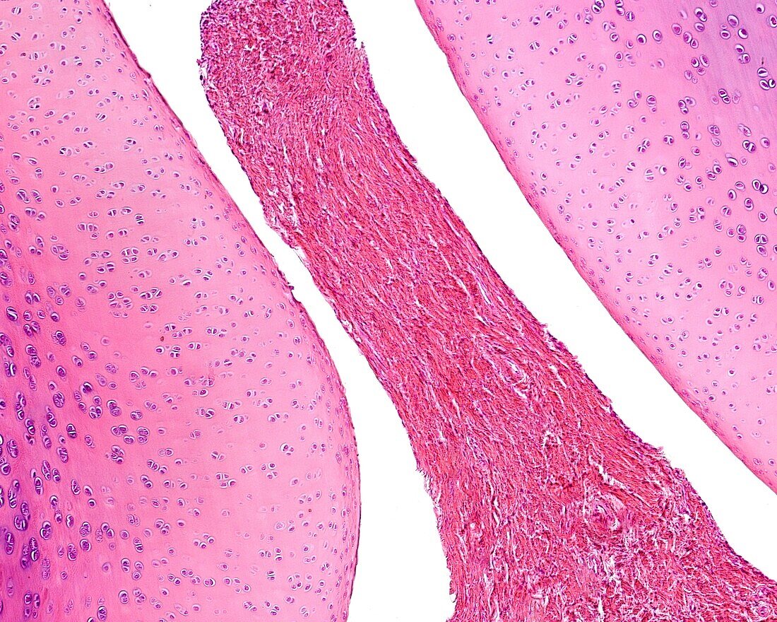
| Light micrograph of a longitudinal section of a knee of a young experimental animal. The epiphysis of the femur (above) is made up of hyaline cartilage and the epiphyseal ossification nucleus has not yet formed. However, in the tibial epiphysis (bottom) the epiphyseal ossification nucleus and the metaphyseal cartilage are evident. Between both articular surfaces there is a narrow articular cavity. At right is the patella and a small wedge of fibrous connective tissue that penetrates the joint cavity corresponding to the meniscus. Below the patella is the attachment of the joint capsule to the periosteum of the tibia. | |
| Licence : | Droits gérés |
| Crédit: | Science Photo Library / JOSE CALVO |
| Taille de l’image : | 3840 px × 3072 px |
| Model Release : | Non requis |
| Property Release : | Non requis |
| Restrictions : | - |
Prix pour cette image À partir de 45 €
Produit vendu
(Calendrier, Carte postale, Carte de vœux, Impression sur textile, Packaging etc)
À partir de 45 €
Usage commercial
(Affichage, Annonce presse, Annonce TV, Carte, Digital - hors rés. sociaux, Digital - rés. sociaux etc)
À partir de 45 €
Éditorial
(Digital, Journal, Livre, Livre pratique, Magazine, Télévision etc)
À partir de 60 €
Usage non-commercial
(Digital - hors rés. sociaux, Digital - rés. sociaux etc)
À partir de 120 €