Compact bone osteons, light micrograph
Numéro d’image : 13452247
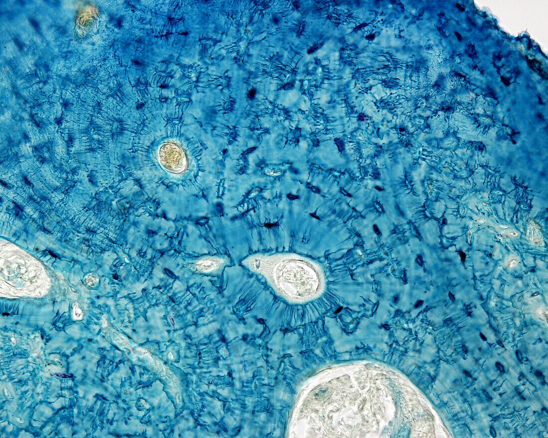
| Light micrograph of compact bone stained with the Schmorl technique, showing osteocytes and their fine processes (blue). There are two osteons, each centred by a rounded space corresponding to the Haversian canal, which is occupied by a small blood vessel. The bone lamellae are arranged around them in concentric layers and the osteocytes are located between them. The numerous extensions that start from the osteocyte soma are striking and interconnect with each other, forming a complex labyrinth. The osteocytes closest to the centre of the osteon orient their processes towards the Haversian canal. | |
| Licence : | Droits gérés |
| Crédit: | Science Photo Library / JOSE CALVO |
| Taille de l’image : | 3840 px × 3072 px |
| Model Release : | Non requis |
| Property Release : | Non requis |
| Restrictions : | - |
Prix pour cette image À partir de 45 €
Produit vendu
(Calendrier, Carte postale, Carte de vœux, Impression sur textile, Packaging etc)
À partir de 45 €
Usage commercial
(Affichage, Annonce presse, Annonce TV, Carte, Digital - hors rés. sociaux, Digital - rés. sociaux etc)
À partir de 45 €
Éditorial
(Digital, Journal, Livre, Livre pratique, Magazine, Télévision etc)
À partir de 60 €
Usage non-commercial
(Digital - hors rés. sociaux, Digital - rés. sociaux etc)
À partir de 120 €