Embryonic bone diaphysis, light micrograph
Numéro d’image : 13452216
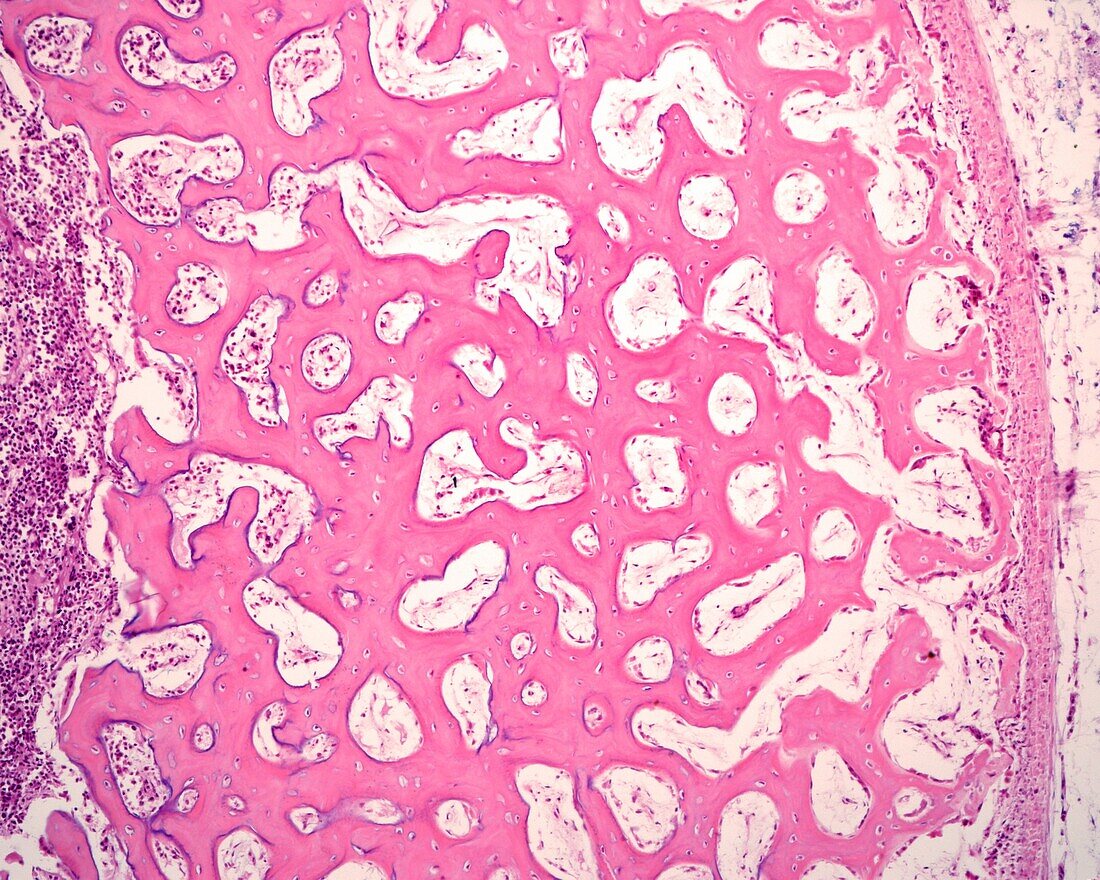
| Peripheral area of the diaphysis of an embryonic bone, light micrograph. The periosteum is at right, showing two layers: an outer, or fibrous layer (pink), and an internal, or osteogenic layer, with a more cellular mesenchyme, where the osteoprogenitor cells are generated. In the more peripheral bone trabeculae, osteoid lines appear formed by osteoblasts that increases the thickness of the diaphyseal cortex. The mean diameter of the trabeculae of immature bone tissue increases from the periosteum to the inside, since, once formed in the periosteum, new layers of bone matrix continue to be attached as time passes. In the internal part, resorption takes place and progressively augments the size of the medullary cavity. | |
| Licence : | Droits gérés |
| Crédit: | Science Photo Library / JOSE CALVO |
| Taille de l’image : | 3840 px × 3072 px |
| Model Release : | Non requis |
| Property Release : | Non requis |
| Restrictions : | - |
Prix pour cette image À partir de 45 €
Produit vendu
(Calendrier, Carte postale, Carte de vœux, Impression sur textile, Packaging etc)
À partir de 45 €
Usage commercial
(Affichage, Annonce presse, Annonce TV, Carte, Digital - hors rés. sociaux, Digital - rés. sociaux etc)
À partir de 45 €
Éditorial
(Digital, Journal, Livre, Livre pratique, Magazine, Télévision etc)
À partir de 60 €
Usage non-commercial
(Digital - hors rés. sociaux, Digital - rés. sociaux etc)
À partir de 120 €
Mots clés
- aucun,
- biologie,
- biologique,
- cartilage,
- chondrocytes,
- croissant,
- développement,
- developpement des os,
- développement du squelette,
- développment osseux,
- épiphyse,
- foetal,
- foetale,
- foetus,
- grandissant,
- histologie,
- histologique,
- matrice du cartilage,
- micrographie optique,
- microscope,
- microscope optique,
- microscopie,
- microscopie optique,
- normal,
- os,
- ossification,
- personne,
- sclérose