Simple columnar epithelium, light micrograph
Numéro d’image : 13452192
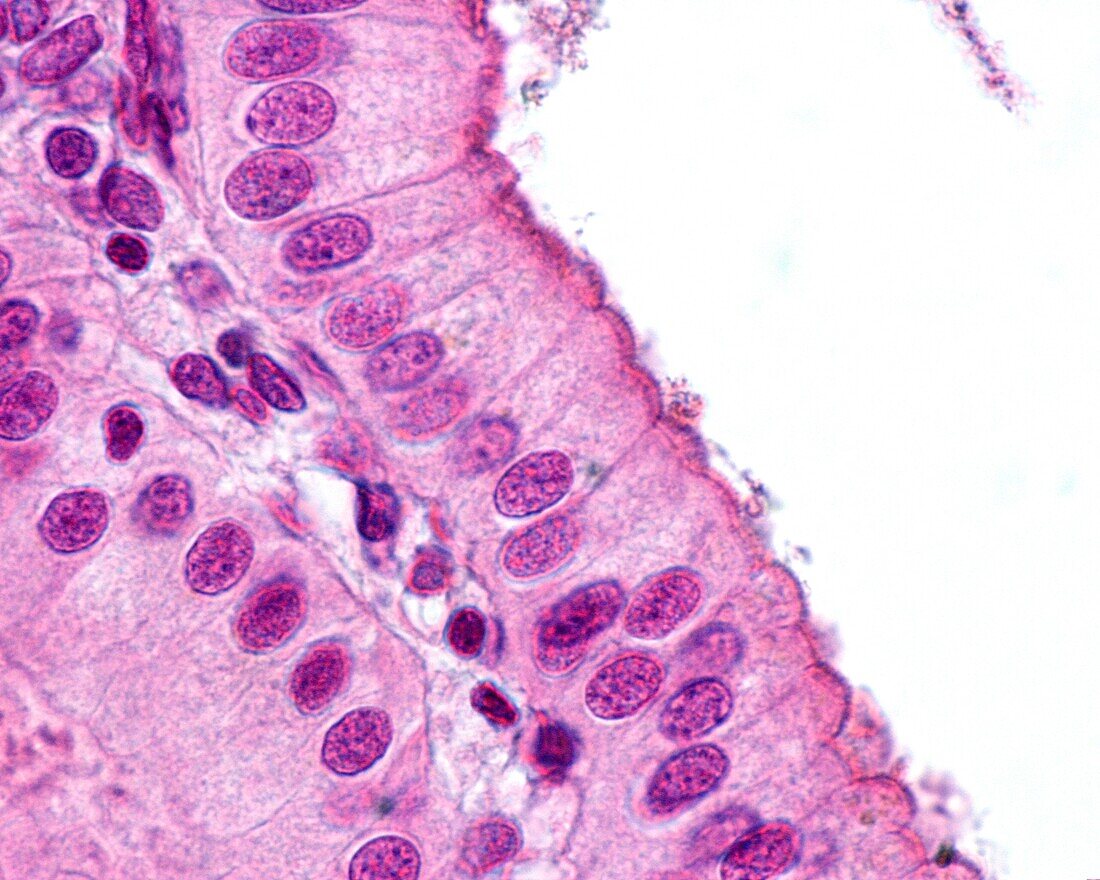
| Light micrograph of gallbladder epithelium. At centre is a fold of the mucous layer, providing a thin axis of lamina propria occupied mainly by blood vessels. Overlying the lamina propria axis is a simple columnar epithelium with nuclei located in the basal third. At the apical border, the cells show a density that corresponds to a striated plate, less developed than in the intestine. The dense nuclei located near the base of the epithelium are lymphocytes. | |
| Licence : | Droits gérés |
| Crédit: | Science Photo Library / JOSE CALVO |
| Taille de l’image : | 3840 px × 3072 px |
| Model Release : | Non requis |
| Property Release : | Non requis |
| Restrictions : | - |
Prix pour cette image À partir de 45 €
Produit vendu
(Calendrier, Carte postale, Carte de vœux, Impression sur textile, Packaging etc)
À partir de 45 €
Usage commercial
(Affichage, Annonce presse, Annonce TV, Carte, Digital - hors rés. sociaux, Digital - rés. sociaux etc)
À partir de 45 €
Éditorial
(Digital, Journal, Livre, Livre pratique, Magazine, Télévision etc)
À partir de 60 €
Usage non-commercial
(Digital - hors rés. sociaux, Digital - rés. sociaux etc)
À partir de 120 €
Mots clés
- aucun,
- biologie,
- biologique,
- corps humain,
- cylindre simple,
- epithelium,
- épithélium,
- épithélium cylindrique,
- foie,
- histologie,
- histologique,
- lamina propria,
- micrographie,
- micrographie optique,
- microscope,
- microscope optique,
- microscopie,
- microscopie optique,
- microscopique,
- mucosa,
- muqueuse,
- personne,
- scientifique,
- système digestif,
- vésicule biliaire