Pine needle, LM
Numéro d’image : 13445253
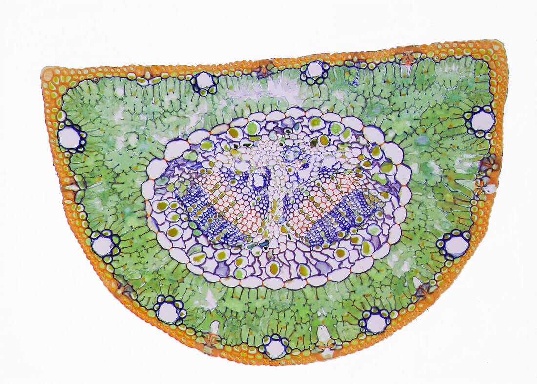
| Section through a pine needle. Light micrograph (LM) of a section through the needle (leaf) of a pine tree, Pinus sp. The centre of the needle is occupied by two vascular bundles (butterfly-shaped region, centre), each one made up of xylem (orange) and phloem (blue) tissue. These are surrounded by a thick pericycle (large-celled region) and a layer of endodermis (necklace-like ring of large cells). The tissue surrounding the endodermis is known as mesophyll (green and brown speckled layer), which contain the resin ducts (not seen). In the epidermis (surface) are stomata (openings) for gas exchange. Magnification x 20 when printed 10cm wide. | |
| Licence : | Droits gérés |
| Crédit: | Science Photo Library / Gschmeissner, Steve |
| Taille de l’image : | 4940 px × 3538 px |
| Model Release : | Non requis |
| Restrictions : | - |
Prix pour cette image À partir de 45 €
Produit vendu
(Calendrier, Carte postale, Carte de vœux, Impression sur textile, Packaging etc)
À partir de 45 €
Usage commercial
(Affichage, Annonce presse, Annonce TV, Carte, Digital - hors rés. sociaux, Digital - rés. sociaux etc)
À partir de 45 €
Éditorial
(Digital, Journal, Livre, Livre pratique, Magazine, Télévision etc)
À partir de 60 €
Usage non-commercial
(Digital - hors rés. sociaux, Digital - rés. sociaux etc)
À partir de 120 €
Mots clés
- aiguille de pin,
- aiguilles de pin,
- biologie,
- biologique,
- botanique,
- catégorie,
- chloroplastes,
- colonial,
- cortex,
- coupe,
- dicotylédonne,
- divisé,
- endoderme,
- endodermis,
- épiderme,
- faisceau vasculaire,
- flore,
- mésophylle,
- micrographie optique,
- microscope optique,
- microscopie optique,
- moelle,
- nature,
- partie,
- phloème,
- pinus,
- plante,
- section,
- tige,
- xylème