Cerebellar cortex Purkinje cells, light micrograph
Numéro d’image : 13416595
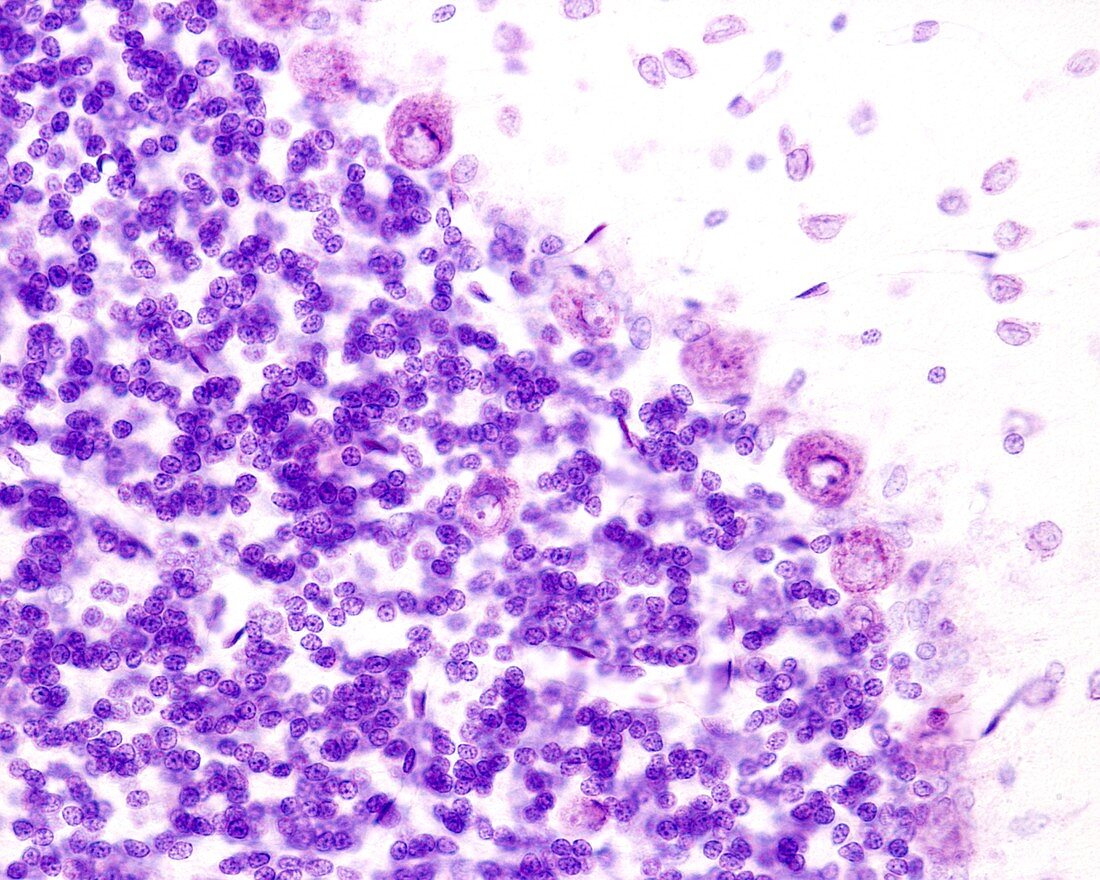
| Light micrograph of the cerebellar cortex showing Purkinje cells located between the molecular and granular layers. A Golgi cell can be seen in the granular layer. Cresyl violet stain. | |
| Licence : | Droits gérés |
| Crédit: | Science Photo Library / JOSE CALVO |
| Taille de l’image : | 3840 px × 3072 px |
| Model Release : | Non requis |
| Property Release : | Non requis |
| Restrictions : | - |
Prix pour cette image À partir de 45 €
Produit vendu
(Calendrier, Carte postale, Carte de vœux, Impression sur textile, Packaging etc)
À partir de 45 €
Usage commercial
(Affichage, Annonce presse, Annonce TV, Carte, Digital - hors rés. sociaux, Digital - rés. sociaux etc)
À partir de 45 €
Éditorial
(Digital, Journal, Livre, Livre pratique, Magazine, Télévision etc)
À partir de 60 €
Usage non-commercial
(Digital - hors rés. sociaux, Digital - rés. sociaux etc)
À partir de 120 €
Mots clés
- aucun,
- cérébelleux,
- cerebellum,
- cervelet,
- cortex,
- éosine,
- fibre,
- granulaire,
- hématoxyline,
- histologie,
- histologique,
- léger,
- matière grise,
- micrographie optique,
- microscope,
- microscope optique,
- microscopie,
- microscopie optique,
- moléculaire,
- myélinisé,
- nerf,
- nerveux,
- neurohistologie,
- neurologie,
- personne,
- S.N.C.,
- SNC,
- système nerveux central