Actinic keratosis, light micrograph
Numéro d’image : 13416568
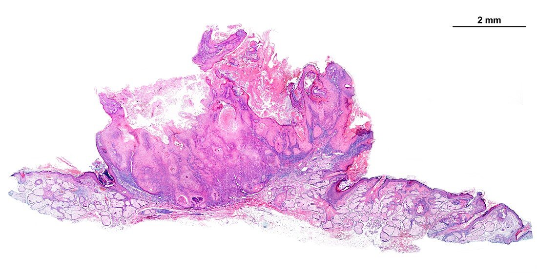
| Light micrograph showing a large lesion of actinic, or senile, keratosis manifested as a skin wart. Visible changes are epidermal thickening, hyperkeratosis and lymphocyte infiltrates in the upper dermis beneath the epidermis. Normal skin can be seen on both sides of the lesion. The scale bar corresponds to 2 mm. | |
| Licence : | Droits gérés |
| Crédit: | Science Photo Library / JOSE CALVO |
| Taille de l’image : | 4911 px × 2500 px |
| Model Release : | Non requis |
| Property Release : | Non requis |
| Restrictions : | - |
Prix pour cette image À partir de 45 €
Produit vendu
(Calendrier, Carte postale, Carte de vœux, Impression sur textile, Packaging etc)
À partir de 45 €
Usage commercial
(Affichage, Annonce presse, Annonce TV, Carte, Digital - hors rés. sociaux, Digital - rés. sociaux etc)
À partir de 45 €
Éditorial
(Digital, Journal, Livre, Livre pratique, Magazine, Télévision etc)
À partir de 60 €
Usage non-commercial
(Digital - hors rés. sociaux, Digital - rés. sociaux etc)
À partir de 120 €