Endochondral ossification, light micrograph
Numéro d’image : 13368800
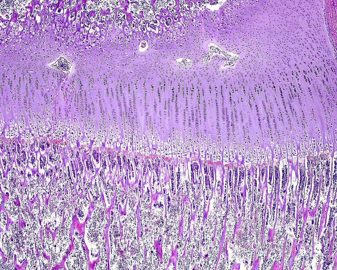
| Light micrograph of the epiphyseal growth plate of a developing long bone. The epiphyseal ossification centre is at top. The epiphyseal cartilage shows the following layers, from top to bottom: resting hyaline cartilage, zone of proliferation, zone of hypertrophy with large lacunae, zone of calcification (not visible as such, but can be identified by the degeneration of the chondrocytes leaving empty cartilaginous lacunae), and an ossification zone of compact appearance, where there is an invasion of blood vessels and osteogenic cells. | |
| Licence : | Droits gérés |
| Crédit: | Science Photo Library / JOSE CALVO |
| Taille de l’image : | 3840 px × 3072 px |
| Model Release : | Non requis |
| Property Release : | Non requis |
| Restrictions : | - |
Prix pour cette image À partir de 45 €
Produit vendu
(Calendrier, Carte postale, Carte de vœux, Impression sur textile, Packaging etc)
À partir de 45 €
Usage commercial
(Affichage, Annonce presse, Annonce TV, Carte, Digital - hors rés. sociaux, Digital - rés. sociaux etc)
À partir de 45 €
Éditorial
(Digital, Journal, Livre, Livre pratique, Magazine, Télévision etc)
À partir de 60 €
Usage non-commercial
(Digital - hors rés. sociaux, Digital - rés. sociaux etc)
À partir de 120 €