Fallopian tube
Numéro d’image : 13356043
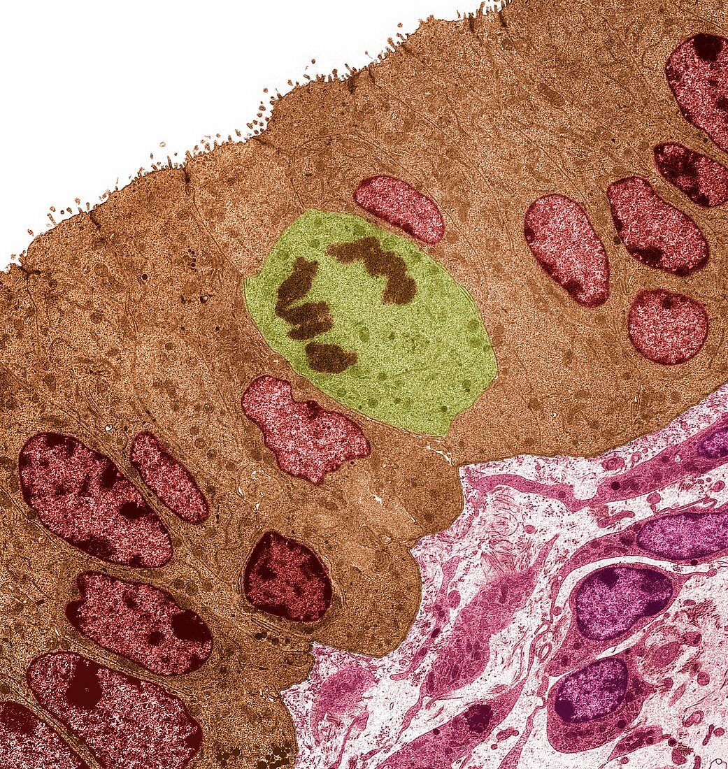
| Fallopian tube, coloured transmission electron micrograph (TEM). Section through columnar epithelium from a fallopian tube during the secretory phase of the endometrial cycle when the ciliated cells are lacking. Large prominent nuclei are present (red) with microvilli visible at the cell surface (top). A mitotic cell is seen center. Connective tissue is seen at the bottom right of the image. Magnification: x1000 when printed at 10 centimetres wide. | |
| Licence : | Droits gérés |
| Crédit: | Science Photo Library / Gschmeissner, Steve |
| Taille de l’image : | 4572 px × 4829 px |
| Model Release : | Non requis |
| Restrictions : | - |
Prix pour cette image À partir de 45 €
Produit vendu
(Calendrier, Carte postale, Carte de vœux, Impression sur textile, Packaging etc)
À partir de 45 €
Usage commercial
(Affichage, Annonce presse, Annonce TV, Carte, Digital - hors rés. sociaux, Digital - rés. sociaux etc)
À partir de 45 €
Éditorial
(Digital, Journal, Livre, Livre pratique, Magazine, Télévision etc)
À partir de 60 €
Usage non-commercial
(Digital - hors rés. sociaux, Digital - rés. sociaux etc)
À partir de 120 €
Mots clés
- anatomie,
- anatomique,
- céllule sécrétrice,
- coloré,
- colorié,
- colorisé,
- corps humain,
- epithelium,
- épithélium,
- féminin,
- féminine,
- humain,
- M.E.T.,
- MET,
- micrographie électronique à transmission,
- microscope électronique à transmission,
- microvillosité,
- oviducte,
- reproduction,
- système reproductif,
- trompe de Fallope,
- trompe utérine,
- trompes de Fallope