Trachea and salivary gland, SEM
Numéro d’image : 13244032
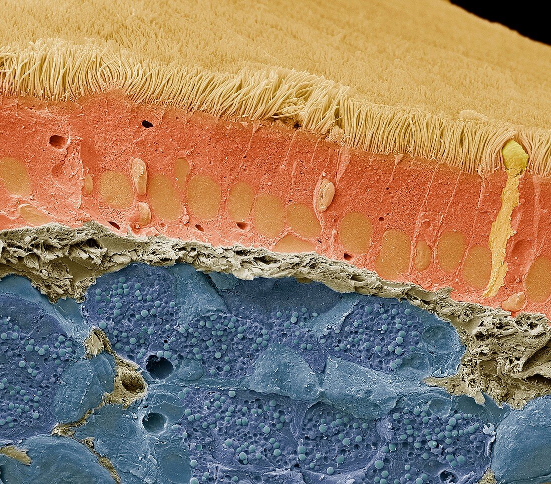
| Trachea and salivary gland. Scanning electron micrograph (SEM) of the trachea and underlying salivary gland. The trachea (top) links the larynx to the lungs. The lining consists of mucus-secreting goblet cells (single cell far right) and epithelial cells that are covered in hair-like cilia. Mucus traps debris, such as dust particles or bacteria, in the inhaled air, while the beating of the cilia moves the mucus and particles upwards out of the respiratory tract. This helps to keep the lungs and airways clear and prevent infection. The salivary gland, shows numerous serous secretory granules (small, spherical), within the serous glands. Serous glands contain serous acini, a grouping of serous cells that secrete a fluid (saliva) containing enzymes such as alpha amylase. Magnification: x1000 when printed at 10cm bacteria | |
| Licence : | Droits gérés |
| Crédit: | Science Photo Library / Gschmeissner, Steve |
| Taille de l’image : | 4572 px × 4016 px |
| Model Release : | Non requis |
| Restrictions : | - |
Prix pour cette image À partir de 45 €
Produit vendu
(Calendrier, Carte postale, Carte de vœux, Impression sur textile, Packaging etc)
À partir de 45 €
Usage commercial
(Affichage, Annonce presse, Annonce TV, Carte, Digital - hors rés. sociaux, Digital - rés. sociaux etc)
À partir de 45 €
Éditorial
(Digital, Journal, Livre, Livre pratique, Magazine, Télévision etc)
À partir de 60 €
Usage non-commercial
(Digital - hors rés. sociaux, Digital - rés. sociaux etc)
À partir de 120 €
Mots clés
- acines,
- acini,
- acinus,
- amylase,
- anatomie,
- anatomique,
- biologie,
- biologique,
- bronches,
- bronchus,
- cellule,
- cils,
- coloré,
- colorié,
- colorisé,
- endoplasmic reticulum,
- granule,
- M.E.B.,
- MEB,
- microscope électronique à balayage,
- noyau,
- nucleus,
- réticulum endoplasmique,
- saliva,
- salivaire,
- salive,
- sécrétion,
- sérosité,
- trachea,
- trachée