Urinary bladder wall, light micrograph
Numéro d’image : 13219530
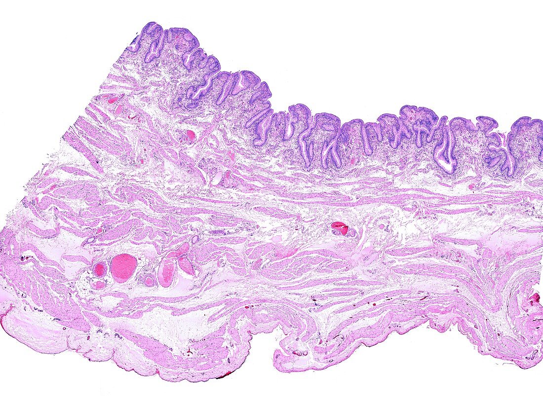
| Low magnification light micrograph of a urinary bladder wall. At the top, lining the lumen of the bladder, is a very folded mucosa layer. Under the mucosae, the muscular layer is formed by smooth muscle cells arranged in thin bundles separated by connective tissue septa with large blood vessels. | |
| Licence : | Droits gérés |
| Crédit: | Science Photo Library / JOSE CALVO |
| Taille de l’image : | 3840 px × 2804 px |
| Model Release : | Non requis |
| Property Release : | Non requis |
| Restrictions : | - |
Prix pour cette image À partir de 45 €
Produit vendu
(Calendrier, Carte postale, Carte de vœux, Impression sur textile, Packaging etc)
À partir de 45 €
Usage commercial
(Affichage, Annonce presse, Annonce TV, Carte, Digital - hors rés. sociaux, Digital - rés. sociaux etc)
À partir de 45 €
Éditorial
(Digital, Journal, Livre, Livre pratique, Magazine, Télévision etc)
À partir de 60 €
Usage non-commercial
(Digital - hors rés. sociaux, Digital - rés. sociaux etc)
À partir de 120 €
Mots clés
- aucun,
- corps humain,
- epithelia,
- épithéliale,
- épithélium,
- épithélium de transition,
- histologie,
- histologique,
- humain,
- micrographie,
- micrographie optique,
- microscope,
- microscope optique,
- microscopie,
- microscopie optique,
- microscopique,
- mucosa,
- muqueuse,
- personne,
- système urinaire,
- tissu conjonctif,
- tissus,
- urinaire,
- urologie,
- urothelium,
- vessie