Cerebral atrophy, MRI scan
Numéro d’image : 13219309
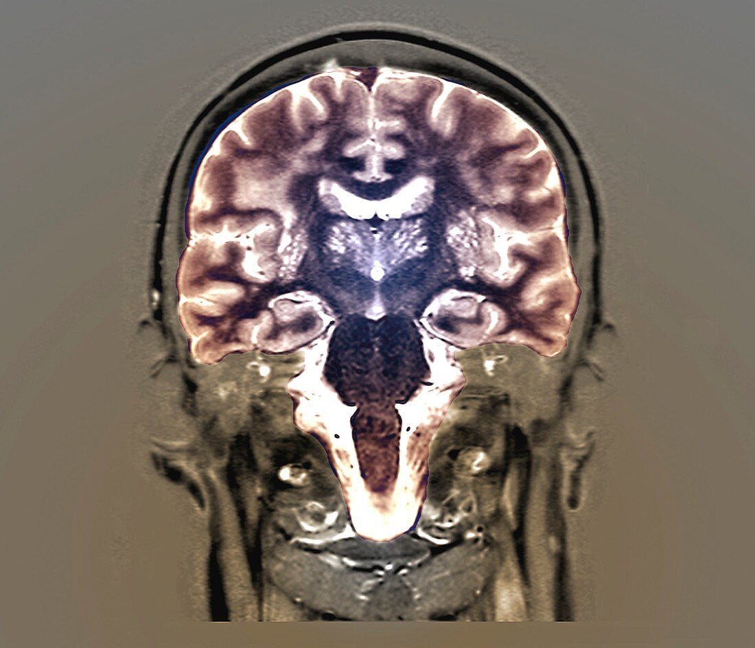
| Coloured magnetic resonance imaging (MRI) scan of a frontal section through the brain of a 74 year old male patient. There are dilated perivascular spaces (white) and nodules (light grey) in the basal ganglia (centre). Perivascular spaces are fluid-filled structures that surround blood vessels. Both of these findings can correspond to cerebral atrophy caused by aging and are also linked to high blood pressure and dementia. | |
| Licence : | Droits gérés |
| Crédit: | Science Photo Library / Zephyr |
| Taille de l’image : | 2854 px × 2449 px |
| Model Release : | Non requis |
| Property Release : | Non requis |
| Restrictions : | - |
Prix pour cette image À partir de 45 €
Produit vendu
(Calendrier, Carte postale, Carte de vœux, Impression sur textile, Packaging etc)
À partir de 45 €
Usage commercial
(Affichage, Annonce presse, Annonce TV, Carte, Digital - hors rés. sociaux, Digital - rés. sociaux etc)
À partir de 45 €
Éditorial
(Digital, Journal, Livre, Livre pratique, Magazine, Télévision etc)
À partir de 60 €
Usage non-commercial
(Digital - hors rés. sociaux, Digital - rés. sociaux etc)
À partir de 120 €
Mots clés
- 70,
- 74,
- années 70,
- anormal,
- atrophie cérébrale,
- aucun,
- cerveau,
- coloré,
- colorié,
- colorisé,
- corps humain,
- diagnostic,
- diagnostique,
- gris,
- homme,
- I.R.M.,
- imagerie par résonance magnétique,
- imagerie par résonnance magnétique,
- IRM,
- malsain,
- masculin,
- médecine,
- médical,
- médicale,
- neurologie,
- neurologique,
- noyaux gris centraux,
- organe,
- personne,
- personnes âgées,
- septante-quatre,
- soixante-quatorze,
- vieillissement,
- vieux