Spinal cord, light micrograph
Numéro d’image : 12987527
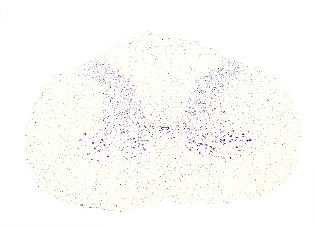
| Cross section of a cervical spinal cord stained with cresyl violet, light micrograph. The central grey matter shows many neuronal cell bodies. In the large anterior horns, big motor neurons (bottom) are seen. | |
| Licence : | Droits gérés |
| Crédit: | Science Photo Library / JOSE CALVO |
| Taille de l’image : | 3840 px × 2749 px |
| Model Release : | Non requis |
| Property Release : | Non requis |
| Restrictions : | - |
Prix pour cette image À partir de 45 €
Produit vendu
(Calendrier, Carte postale, Carte de vœux, Impression sur textile, Packaging etc)
À partir de 45 €
Usage commercial
(Affichage, Annonce presse, Annonce TV, Carte, Digital - hors rés. sociaux, Digital - rés. sociaux etc)
À partir de 45 €
Éditorial
(Digital, Journal, Livre, Livre pratique, Magazine, Télévision etc)
À partir de 60 €
Usage non-commercial
(Digital - hors rés. sociaux, Digital - rés. sociaux etc)
À partir de 120 €
Mots clés
- aucun,
- biologie,
- biologique,
- histologie,
- histologique,
- matière blanche,
- matière grise,
- microscope,
- microscope optique,
- microscopie,
- microscopie optique,
- microscopique,
- moelle épinière,
- moteur,
- nerveux,
- neurohistologie,
- neurologie,
- neurologique,
- neurone,
- neurones,
- personne,
- système,
- tissus,
- vertébral