Intestine microvilli, TEM
Numéro d’image : 12949502
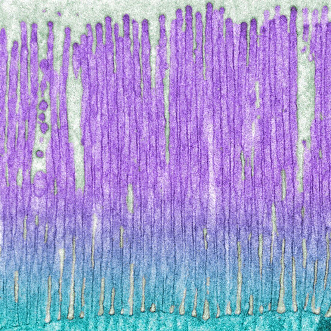
| Intestine microvilli, coloured transmission electron micrograph (TEM). Epithelial cells from the intestine absorb nutrients from digested food. Increasing the surface area of these cells allows for this absorption to be more efficient. The surface area of the cells is increased by the cells being covered in microvilli which are long finger-like projections which form a brush border. Surrounding the microvilli is the glycocalyx. This is a sticky layer comprised of glycoproteins and glycolipids. Magnification: x28000 when printed at 10cm wide. | |
| Licence : | Droits gérés |
| Crédit: | Science Photo Library / Anne Weston, EM STP, the Francis Crick Institute |
| Taille de l’image : | 5315 px × 5315 px |
| Model Release : | Non requis |
| Property Release : | Non requis |
| Restrictions : |
|
Prix pour cette image À partir de 45 €
Produit vendu
(Calendrier, Carte postale, Carte de vœux, Impression sur textile, Packaging etc)
À partir de 45 €
Usage commercial
(Affichage, Annonce presse, Annonce TV, Carte, Digital - hors rés. sociaux, Digital - rés. sociaux etc)
À partir de 45 €
Éditorial
(Digital, Journal, Livre, Livre pratique, Magazine, Télévision etc)
À partir de 60 €
Usage non-commercial
(Digital - hors rés. sociaux, Digital - rés. sociaux etc)
À partir de 120 €
Mots clés
- absorption,
- aucun,
- biologie,
- biologique,
- bordure en brosse,
- cellule,
- coloré,
- colorié,
- colorisé,
- en bonne santé,
- epithelium,
- épithélium,
- glycocalyx,
- histologie,
- histologique,
- intestin,
- intestinal,
- M.E.T.,
- MET,
- micrographie électronique à transmission,
- microscope électronique à transmission,
- microscope électronique en transmission,
- microvillosité,
- microvillus,
- normal,
- personne,
- plycoprotéine,
- sain,
- système digestif