Cartilage, SEM-TEM comparison
Numéro d’image : 12948521
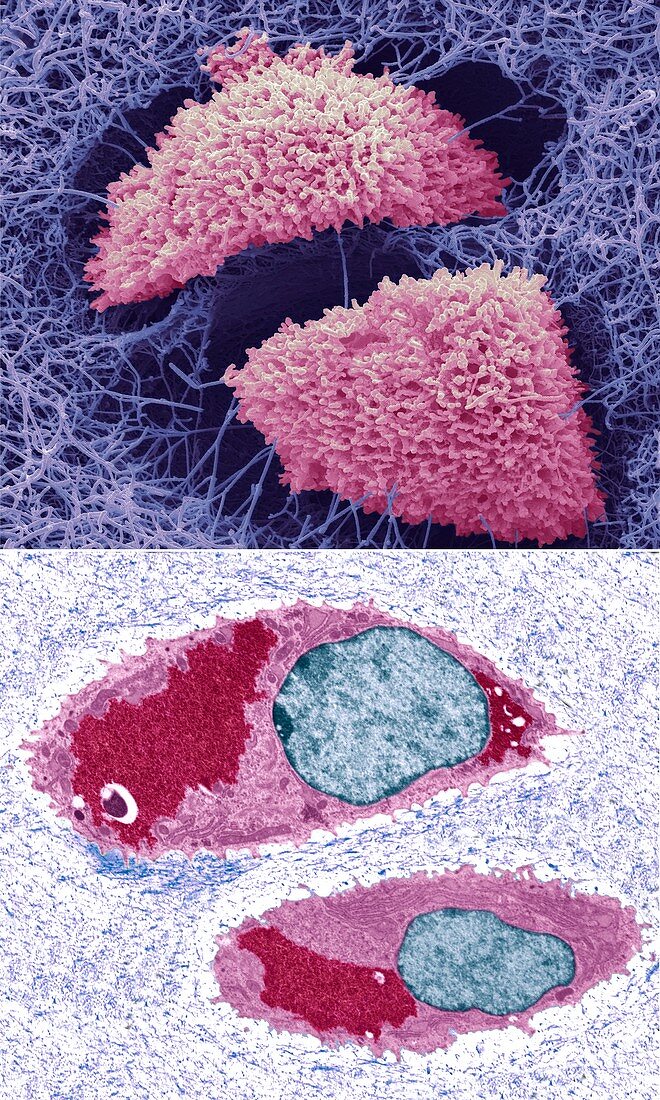
| Comparison between a scanning electron micrograph (SEM, top) and transmission electron micrograph (TEM, bottom) of cartilage cells. A pair of chondrocytes (pink) are at centre. Chondrocytes are the only cells found in cartilage, a connective tissue that provides structural support. The cells produce and maintain the cartilage's extracellular matrix (blue). In the The TEM image the cell nuclei are blue. In their cytoplasm (pink) are collections of glycogen granules (red), which provide the cell with energy. Magnification: x6000 when printed at 10 centimetres wide. For a series of comparisons between SEMs and TEMs see images C047/7006 to C047/7034. | |
| Licence : | Droits gérés |
| Crédit: | Science Photo Library / Gschmeissner, Steve |
| Taille de l’image : | 4572 px × 7620 px |
| Model Release : | Non requis |
| Property Release : | Non requis |
| Restrictions : | - |
Prix pour cette image À partir de 45 €
Produit vendu
(Calendrier, Carte postale, Carte de vœux, Impression sur textile, Packaging etc)
À partir de 45 €
Usage commercial
(Affichage, Annonce presse, Annonce TV, Carte, Digital - hors rés. sociaux, Digital - rés. sociaux etc)
À partir de 45 €
Éditorial
(Digital, Journal, Livre, Livre pratique, Magazine, Télévision etc)
À partir de 60 €
Usage non-commercial
(Digital - hors rés. sociaux, Digital - rés. sociaux etc)
À partir de 120 €
Mots clés
- anatomie,
- anatomique,
- biologie,
- biologique,
- cartilage,
- cellule,
- chondrocytes,
- coloré,
- colorié,
- colorisé,
- comparaison,
- comparé,
- comparer,
- glycogène,
- lacune,
- M.E.B.,
- M.E.T.,
- matrice extracellulaire,
- MEB,
- MET,
- micrographie électronique à transmission,
- microscope électronique à balayage,
- microscope électronique à transmission,
- noyau,
- nucleus,
- organites,
- séquence,
- séries,
- structurel,
- tissu conjonctif