Ovarian secondary follicle,light micrograph
Numéro d’image : 12894118
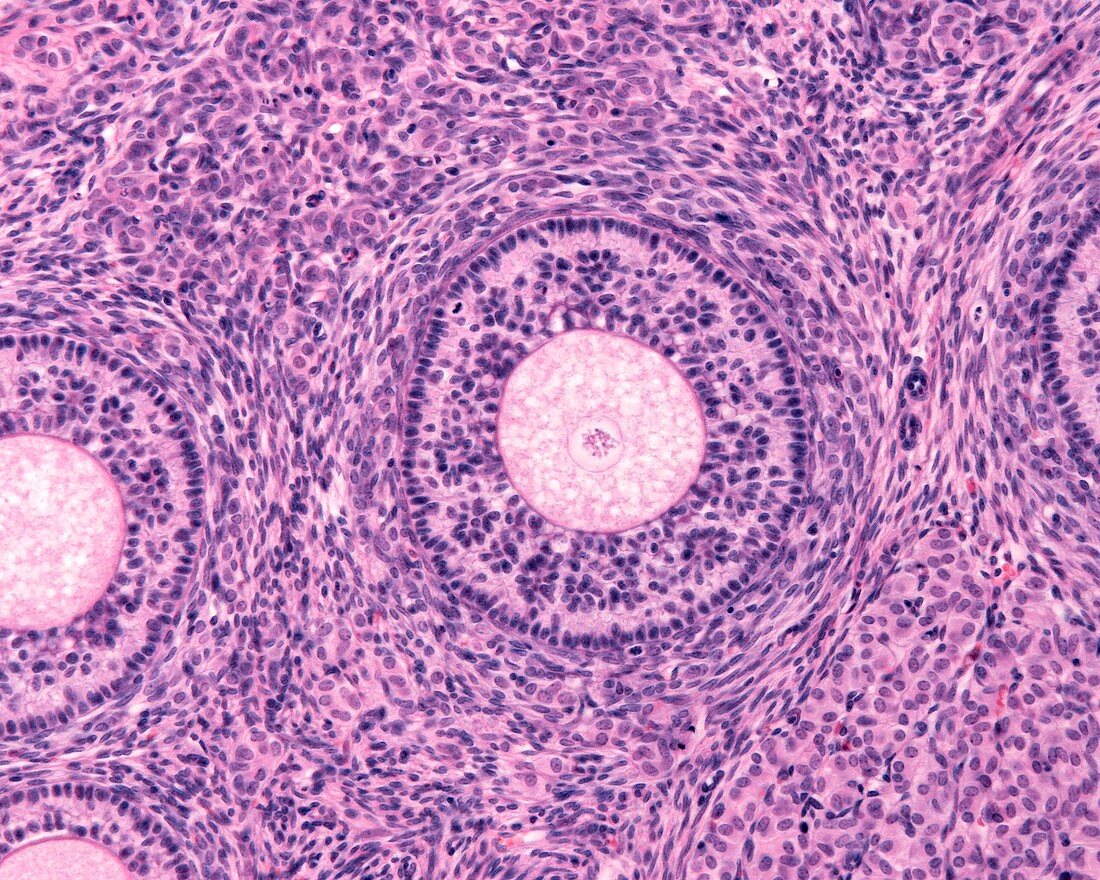
| Light micrograph of a secondary follicle showing oocyte,zona pellucida,and granulosa cells organized in several layers and showing mitosis. Outside the follicle can be seen both thecas,interna and externa. | |
| Licence : | Droits gérés |
| Crédit: | Science Photo Library / JOSE CALVO |
| Taille de l’image : | 4674 px × 3739 px |
| Model Release : | Non requis |
| Property Release : | Non requis |
| Restrictions : | - |
Prix pour cette image À partir de 45 €
Produit vendu
(Calendrier, Carte postale, Carte de vœux, Impression sur textile, Packaging etc)
À partir de 45 €
Usage commercial
(Affichage, Annonce presse, Annonce TV, Carte, Digital - hors rés. sociaux, Digital - rés. sociaux etc)
À partir de 45 €
Éditorial
(Digital, Journal, Livre, Livre pratique, Magazine, Télévision etc)
À partir de 60 €
Usage non-commercial
(Digital - hors rés. sociaux, Digital - rés. sociaux etc)
À partir de 120 €
Mots clés
- anatomie microscopique,
- cellule,
- éosine,
- féminin,
- féminine,
- follicule,
- follicule ovarien,
- granulosa,
- hématoxyline,
- histologie,
- histologique,
- membrane pellucide,
- micrographie,
- microscope,
- microscopie,
- microscopique,
- mitose,
- originel,
- ovaire,
- ovarien,
- ovocyte,
- primaire,
- primordial,
- reproductif,
- secondaire,
- système reproducteur féminin,
- tissus,
- zona pellicida,
- zona pellucida,
- zone pellucide