Subacute Sclerosing Panencephalitis
Numéro d’image : 12652237
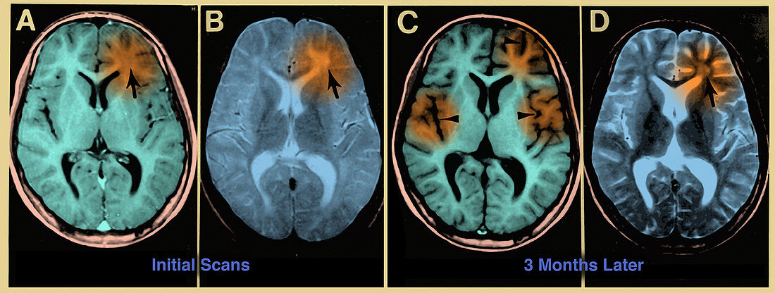
| Subacute sclerosing panencephalitis (a complication of measles infection). Figure 1. MRI scans of the brain at the time of presentation in the neurology clinic (A and B) and 3 months later (C and D). Panels A and C are T1-weighted images, B and D are T2-weighted images. The initial MRI scan (A and B) reveals a focal abnormality in the subcortical white matter of the left frontal lobe, consisting of a hypointense signal on the T1-weighted image (arrow in A) and a hyperintense signal on the T2-weighted image (arrow in B). In the follow-up scan, the focal abnormality in the left frontal lobe is less obvious than previously (arrow in D), but advanced and diffuse cortical atrophy is present, signified by the ventriculomegaly and markedly enlarged sulci (arrowheads in C). | |
| Licence : | Droits gérés |
| Crédit: | Science Photo Library / Science Source |
| Taille de l’image : | 5287 px × 2000 px |
| Model Release : | Non requis |
| Property Release : | Non requis |
| Restrictions : | - |
Prix pour cette image À partir de 45 €
Produit vendu
(Calendrier, Carte postale, Carte de vœux, Impression sur textile, Packaging etc)
À partir de 45 €
Usage commercial
(Affichage, Annonce presse, Annonce TV, Carte, Digital - hors rés. sociaux, Digital - rés. sociaux etc)
À partir de 45 €
Éditorial
(Digital, Journal, Livre, Livre pratique, Magazine, Télévision etc)
À partir de 60 €
Usage non-commercial
(Digital - hors rés. sociaux, Digital - rés. sociaux etc)
À partir de 120 €
Mots clés
- amélioration,
- amélioré,
- augmenté,
- cerveau,
- complication,
- coupe transversale,
- encéphalite,
- encephalitis,
- I.R.M.,
- imagerie médicale,
- imagerie par résonance magnétique,
- imagerie par résonnance magnétique,
- inflammation,
- IRM,
- lobe frontal,
- médecine,
- médical,
- médicale,
- médicinal,
- renforcé,
- rougeole,
- scanner,
- scanner du cerveau