Physical acinar, ductal, and insular interaction
Numéro d’image : 12650621
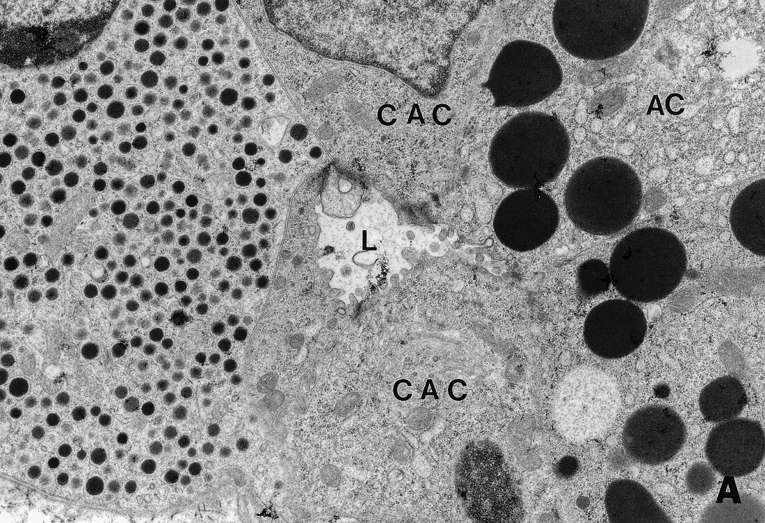
| Transmission electron microscope image of cellular junction between acinar, (AC) cells, having zymogen granules, centroacinar cells (CAC) and islet cell (left) filled with endocrine granules. These cells are attached to each other by junctional complexes as seen as dark areas around the lumen (L), TEM x 2, 200. | |
| Licence : | Droits gérés |
| Crédit: | Science Photo Library / Science Source / Nature's Faces |
| Taille de l’image : | 2945 px × 2017 px |
| Model Release : | Non requis |
| Property Release : | Non requis |
| Restrictions : | - |
Prix pour cette image À partir de 45 €
Produit vendu
(Calendrier, Carte postale, Carte de vœux, Impression sur textile, Packaging etc)
À partir de 45 €
Usage commercial
(Affichage, Annonce presse, Annonce TV, Carte, Digital - hors rés. sociaux, Digital - rés. sociaux etc)
À partir de 45 €
Éditorial
(Digital, Journal, Livre, Livre pratique, Magazine, Télévision etc)
À partir de 60 €
Usage non-commercial
(Digital - hors rés. sociaux, Digital - rés. sociaux etc)
À partir de 120 €
Mots clés
- acineux,
- canalaire,
- cellulaire,
- cellule,
- cellules,
- ductal,
- endocrine,
- granule,
- granule zymogène,
- granules,
- granulés,
- îlot,
- lumen,
- lumière,
- M.E.,
- M.E.T.,
- ME,
- MET,
- micrographie électronique,
- micrographie électronique à transmission,
- microscope électronique,
- microscope électronique à transmission,
- microscope électronique en transmission,
- microscopie électronique,
- pancréas,
- pancréatique,
- proenzyme,
- zymogène