Liver cell degeneration, LM
Numéro d’image : 12648502
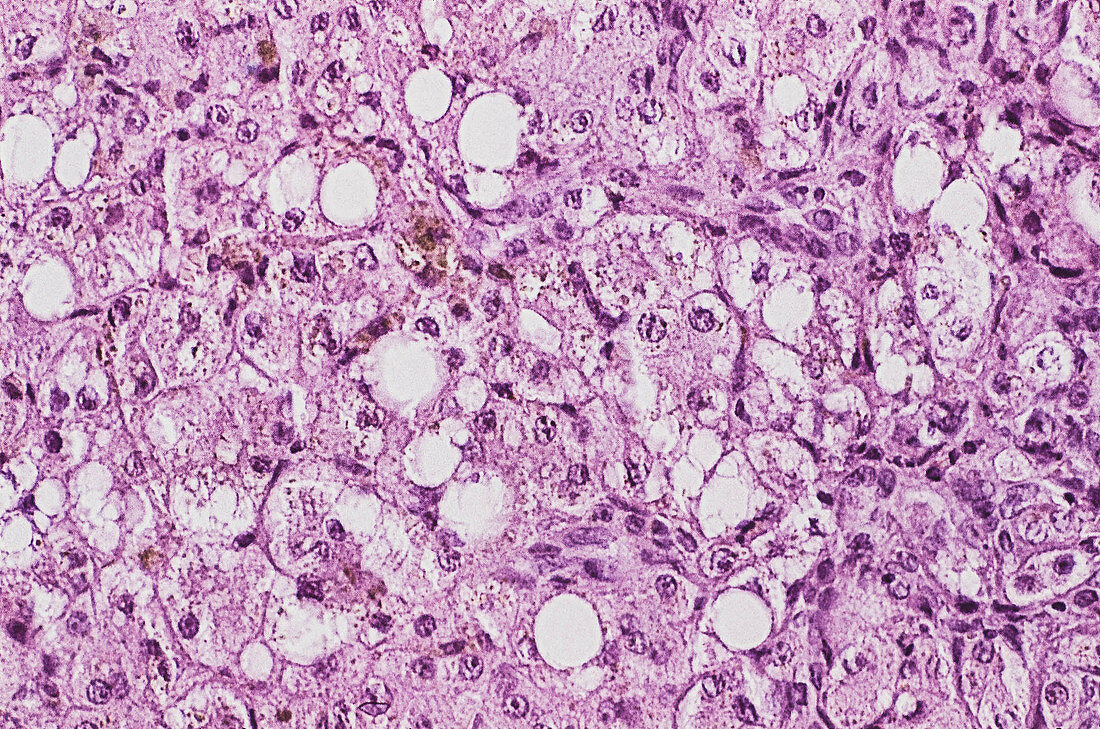
| Histological microscopical findings of the liver showing degeneration of many cells appearing as holes that present fat and brownish pigment indicating iron pigment (hemosiderin) due to bleeding. H&E stain, 40x magnification. | |
| Licence : | Droits gérés |
| Crédit: | Science Photo Library / Science Source / Nature's Faces |
| Taille de l’image : | 2168 px × 1437 px |
| Model Release : | Non requis |
| Property Release : | Non requis |
| Restrictions : | - |
Prix pour cette image À partir de 45 €
Produit vendu
(Calendrier, Carte postale, Carte de vœux, Impression sur textile, Packaging etc)
À partir de 45 €
Usage commercial
(Affichage, Annonce presse, Annonce TV, Carte, Digital - hors rés. sociaux, Digital - rés. sociaux etc)
À partir de 45 €
Éditorial
(Digital, Journal, Livre, Livre pratique, Magazine, Télévision etc)
À partir de 60 €
Usage non-commercial
(Digital - hors rés. sociaux, Digital - rés. sociaux etc)
À partir de 120 €
Mots clés
- anomalie,
- anormal,
- anormale,
- anormalité,
- cellules du foie,
- dégénération,
- dégénérescence,
- dégradation,
- désordre,
- état,
- histologie,
- histologique,
- malade,
- maladie,
- micrographie,
- micrographie optique,
- microscope,
- microscope optique,
- microscope photonique,
- microscopie,
- microscopie optique,
- microscopie photonique,
- nécrose,
- necrosis,
- pathologie,
- pathologique,
- trouble