MRI of Arterial Venous Fistulas 3
Numéro d’image : 12648192
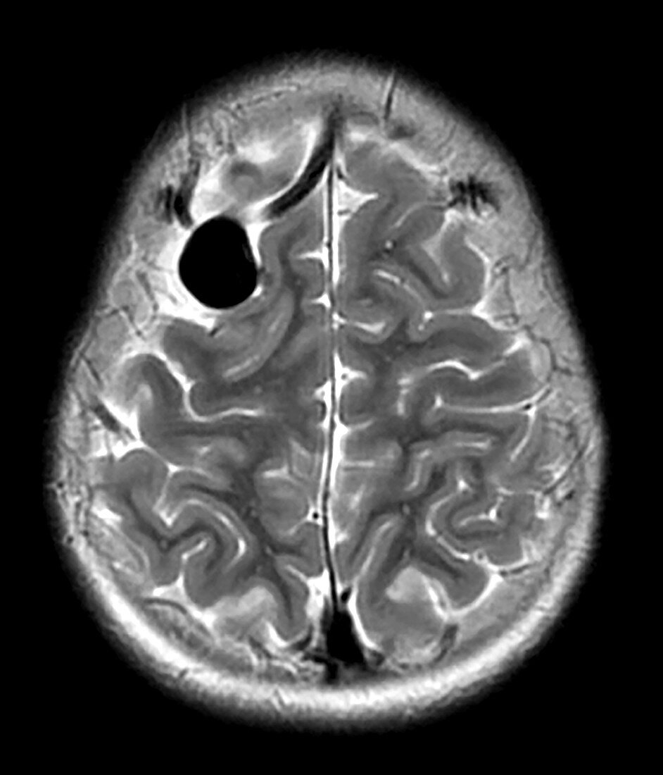
| This axial (cross sectional) T2 weighted MR image shows an enlarged vascular channel in the frontal lobe on viewers left reflecting an abnormal connection between an artery and vein (with abscess of the intervening capillary bed). This is called an AV (arterial venous) fistula. | |
| Licence : | Droits gérés |
| Crédit: | Science Photo Library / Science Source / Living Art Enterprises, LLC |
| Taille de l’image : | 3900 px × 4554 px |
| Model Release : | Non requis |
| Property Release : | Non requis |
| Restrictions : | - |
Prix pour cette image À partir de 45 €
Produit vendu
(Calendrier, Carte postale, Carte de vœux, Impression sur textile, Packaging etc)
À partir de 45 €
Usage commercial
(Affichage, Annonce presse, Annonce TV, Carte, Digital - hors rés. sociaux, Digital - rés. sociaux etc)
À partir de 45 €
Éditorial
(Digital, Journal, Livre, Livre pratique, Magazine, Télévision etc)
À partir de 60 €
Usage non-commercial
(Digital - hors rés. sociaux, Digital - rés. sociaux etc)
À partir de 120 €