Pterional Meningioma on MRI
Numéro d’image : 12646862
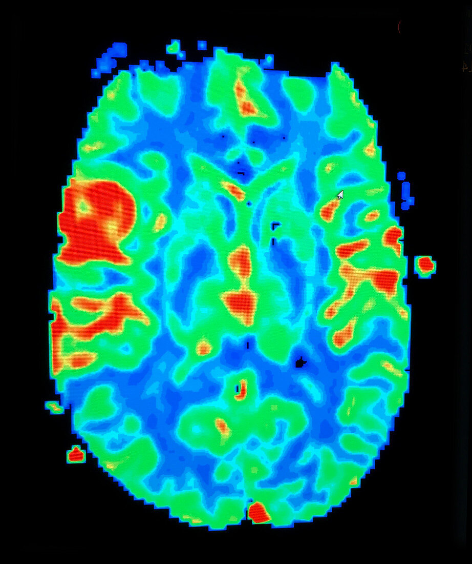
| This axial (cross sectional) image from an MR perfusion (DSC, dynamic susceptibility contrast technique) shows increased CBV (cerebral blood volume) and CBF (cerebral blood flow) of a meningioma in the region of the right pterion and right sylvian fissure. This appears as the area in red on the viewers left. | |
| Licence : | Droits gérés |
| Crédit: | Science Photo Library / Science Source / Living Art Enterprises, LLC |
| Taille de l’image : | 3900 px × 4651 px |
| Model Release : | Non requis |
| Property Release : | Non requis |
| Restrictions : | - |
Prix pour cette image À partir de 45 €
Produit vendu
(Calendrier, Carte postale, Carte de vœux, Impression sur textile, Packaging etc)
À partir de 45 €
Usage commercial
(Affichage, Annonce presse, Annonce TV, Carte, Digital - hors rés. sociaux, Digital - rés. sociaux etc)
À partir de 45 €
Éditorial
(Digital, Journal, Livre, Livre pratique, Magazine, Télévision etc)
À partir de 60 €
Usage non-commercial
(Digital - hors rés. sociaux, Digital - rés. sociaux etc)
À partir de 120 €