Hereditary hemochromatosis, light micrograph
Numéro d’image : 12645087
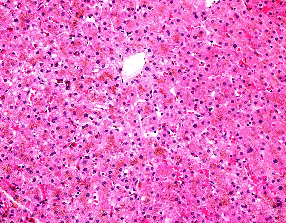
| Light micrograph of a section of liver from a patient with classic HFE-linked hemochromatosis (hemochromatosis type 1). There is accumulation of hemosiderin granules within hepatocytes in lysosomal location. The iron storage shows a gradient from portal to central zones. With progressive iron deposition, there is portal fibrosis with expansion of portal tracts, ultimately resulting in diffuse micronodular cirrhosis. | |
| Licence : | Droits gérés |
| Crédit: | Science Photo Library / WEBPATHOLOGY |
| Taille de l’image : | 4730 px × 3695 px |
| Model Release : | Non requis |
| Property Release : | Non requis |
| Restrictions : | - |
Prix pour cette image À partir de 45 €
Produit vendu
(Calendrier, Carte postale, Carte de vœux, Impression sur textile, Packaging etc)
À partir de 45 €
Usage commercial
(Affichage, Annonce presse, Annonce TV, Carte, Digital - hors rés. sociaux, Digital - rés. sociaux etc)
À partir de 45 €
Éditorial
(Digital, Journal, Livre, Livre pratique, Magazine, Télévision etc)
À partir de 60 €
Usage non-commercial
(Digital - hors rés. sociaux, Digital - rés. sociaux etc)
À partir de 120 €
Mots clés
- cirrhose,
- cirrhosis,
- corps humain,
- désordre,
- état,
- foie,
- gastro-entérologue,
- gastroentérologie,
- gastroentérologue,
- génétique,
- hématochromatose,
- hémochromatose,
- hépatique,
- héréditaire,
- histologie,
- histologique,
- histopathologie,
- histopathologique,
- maladie,
- médecine,
- médical,
- médicale,
- microscope optique,
- microscopie optique,
- pathologie,
- pathologique,
- trouble