Nasal epithelium, SEM
Numéro d’image : 12643021
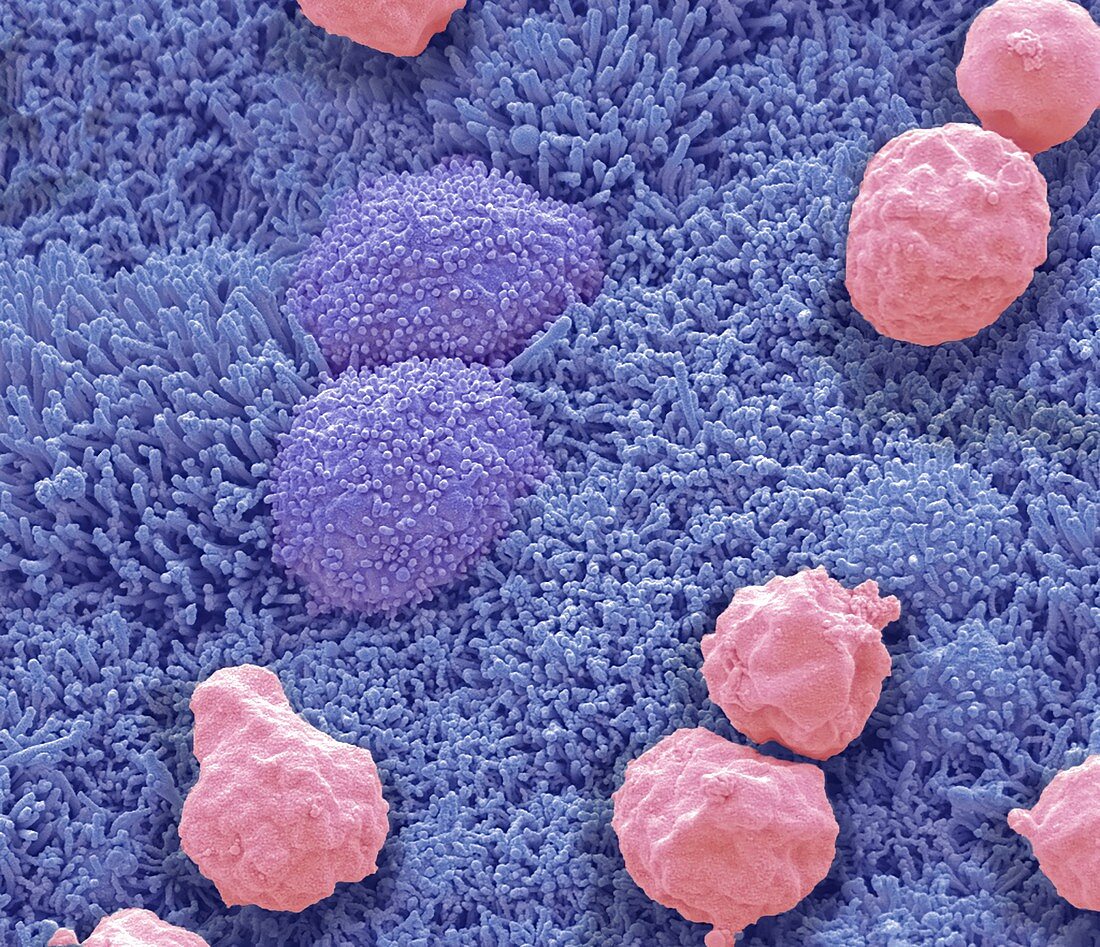
| Nasal epithelium, coloured scanning electron micrograph (SEM). Shown here are squamous nasal epithelial cells (blue/purple) and mucous (pink). The epithelial cell surfaces are covered with tiny microvilli that increase the cell surface area. The microvilli likely aid in localization of foreign debris (coming from the nose) and detection by immune cells. Mucus, secreted by cells in the epithelial lining, traps foreign objects such as bacteria, preventing them from entering the lungs. Magnification: x4000 when printed at 10 centimetres wide. | |
| Licence : | Droits gérés |
| Crédit: | Science Photo Library / Gschmeissner, Steve |
| Taille de l’image : | 4572 px × 3938 px |
| Model Release : | Non requis |
| Property Release : | Non requis |
| Restrictions : | - |
Prix pour cette image À partir de 45 €
Produit vendu
(Calendrier, Carte postale, Carte de vœux, Impression sur textile, Packaging etc)
À partir de 45 €
Usage commercial
(Affichage, Annonce presse, Annonce TV, Carte, Digital - hors rés. sociaux, Digital - rés. sociaux etc)
À partir de 45 €
Éditorial
(Digital, Journal, Livre, Livre pratique, Magazine, Télévision etc)
À partir de 60 €
Usage non-commercial
(Digital - hors rés. sociaux, Digital - rés. sociaux etc)
À partir de 120 €
Mots clés
- anatomie,
- anatomique,
- appareil,
- bacteria,
- bactérie,
- biologie,
- biologique,
- cellule,
- cil,
- coloré,
- colorié,
- colorisé,
- doublure,
- en bonne santé,
- epithelia,
- epithelium,
- épithélium,
- garniture,
- histologie,
- histologique,
- interne,
- M.E.B.,
- MEB,
- microscope électronique à balayage,
- muqueuse,
- muqueux,
- nasal,
- nez,
- normal,
- pulmonaire,
- respiration,
- revêtment,
- sain,
- surface,
- système respiratoire,
- voie