Lumbar Myelogram with Instrumentation, X-ray
Numéro d’image : 12642211
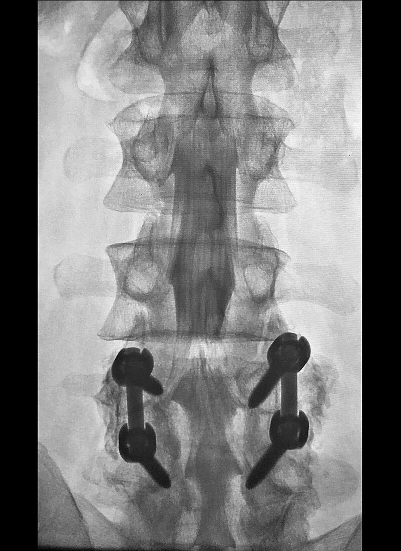
| This frontal x-ray of the lumbar spine during a myelogram shows L4 and L5 pedicle screws and grade 1 spondylolisthesis of L4 over L5. The thecal sac is filled with contrast which is injected after a spinal tap. The string like densities inside of the contrast filled thecal sac are the nerve roots of the cauda equina (so named because they resemble a horse0s tail). | |
| Licence : | Droits gérés |
| Crédit: | Science Photo Library / Living Art Enterprises, LLC |
| Taille de l’image : | 3900 px × 5354 px |
| Model Release : | Non requis |
| Property Release : | Non requis |
| Restrictions : | - |
Prix pour cette image À partir de 45 €
Produit vendu
(Calendrier, Carte postale, Carte de vœux, Impression sur textile, Packaging etc)
À partir de 45 €
Usage commercial
(Affichage, Annonce presse, Annonce TV, Carte, Digital - hors rés. sociaux, Digital - rés. sociaux etc)
À partir de 45 €
Éditorial
(Digital, Journal, Livre, Livre pratique, Magazine, Télévision etc)
À partir de 60 €
Usage non-commercial
(Digital - hors rés. sociaux, Digital - rés. sociaux etc)
À partir de 120 €
Mots clés
- anatomie,
- anatomique,
- anomalie,
- anomalies,
- anormal,
- anormale,
- anormalité,
- désordre,
- diagnostic,
- diagnostique,
- état,
- frontal,
- lumbago,
- maladie,
- malformations,
- médecine,
- médical,
- médicale,
- myélographie,
- rachis lombaire,
- radiculopathie lombaire,
- radiographie,
- radiologie,
- représentation,
- scanner,
- spondylolisthésis,
- trouble,
- vis pédiculaire,
- vis pédonculaire