Brain, MRI
Numéro d’image : 12637627
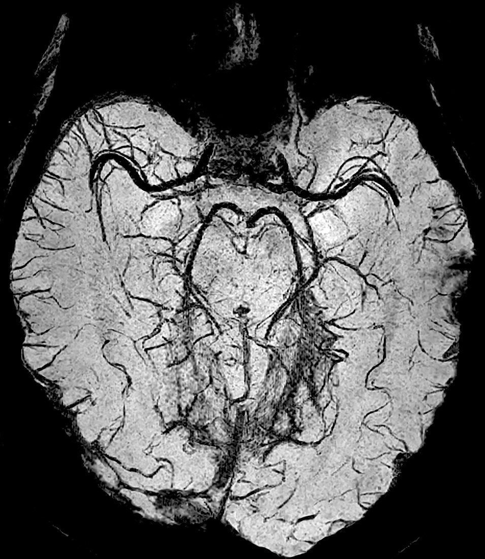
| Normal axial brain SWI (Susceptibility Weighted Imaging) MRI demonstrating the intracranial venous anatomy. There is also visualization of the intracranial arterial system. | |
| Licence : | Droits gérés |
| Crédit: | Science Photo Library / Living Art Enterprises |
| Taille de l’image : | 3900 px × 4493 px |
| Model Release : | Non requis |
| Property Release : | Non requis |
| Restrictions : | - |
Prix pour cette image À partir de 45 €
Produit vendu
(Calendrier, Carte postale, Carte de vœux, Impression sur textile, Packaging etc)
À partir de 45 €
Usage commercial
(Affichage, Annonce presse, Annonce TV, Carte, Digital - hors rés. sociaux, Digital - rés. sociaux etc)
À partir de 45 €
Éditorial
(Digital, Journal, Livre, Livre pratique, Magazine, Télévision etc)
À partir de 60 €
Usage non-commercial
(Digital - hors rés. sociaux, Digital - rés. sociaux etc)
À partir de 120 €
Mots clés
- anatomie,
- anatomique,
- calcium,
- cérébral,
- cerveau,
- fonction,
- fonctionner,
- I.R.M.,
- image ponderée fonction sensibilité,
- imagerie médicale,
- imagerie par résonance magnétique,
- imagerie par résonnance magnétique,
- IRM,
- IRM avancé,
- IRM plus poussé,
- médecine,
- médical,
- médicale,
- normal,
- recherche,
- sang,
- scanner,
- vaisseaux sanguins cérébraux