Subependymoma, MRI
Numéro d’image : 12636820
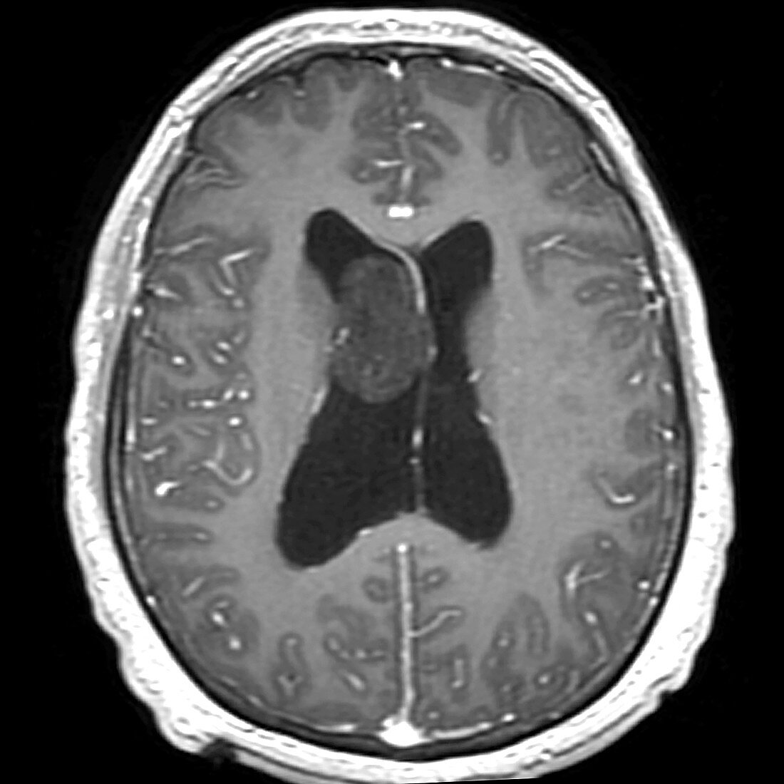
| This axial (cross section) contrast enhanced T1 weighted MRI shows a well circumscribed, non-enhancing mass within the lateral ventricle with an attachment to the septum pellucidum. The mass demonstrates decreased T1 signal with some mild heterogeneity. This is the typical appearance of a Subependymoma, a benign variant of the ependymoma tumours which is often seen as an incidental finding in the elderly. | |
| Licence : | Droits gérés |
| Crédit: | Science Photo Library / Living Art Enterprises |
| Taille de l’image : | 3900 px × 3900 px |
| Model Release : | Non requis |
| Property Release : | Non requis |
| Restrictions : | - |
Prix pour cette image À partir de 45 €
Produit vendu
(Calendrier, Carte postale, Carte de vœux, Impression sur textile, Packaging etc)
À partir de 45 €
Usage commercial
(Affichage, Annonce presse, Annonce TV, Carte, Digital - hors rés. sociaux, Digital - rés. sociaux etc)
À partir de 45 €
Éditorial
(Digital, Journal, Livre, Livre pratique, Magazine, Télévision etc)
À partir de 60 €
Usage non-commercial
(Digital - hors rés. sociaux, Digital - rés. sociaux etc)
À partir de 120 €
Mots clés
- diagnostic,
- diagnostique,
- I.R.M.,
- imagerie médicale,
- imagerie par résonance magnétique,
- imagerie par résonnance magnétique,
- IRM,
- lésion cérébrale,
- masse cérébrale,
- masse du cerveau,
- médecine,
- médical,
- médicale,
- néoplasme du cerveau,
- neurologie,
- neurologique,
- radiographie,
- système nerveux,
- tumeur cérébrale bénigne,
- tumeurs cérébrales