Neurofibromatosis type I (NF1), MRI
Numéro d’image : 12636813
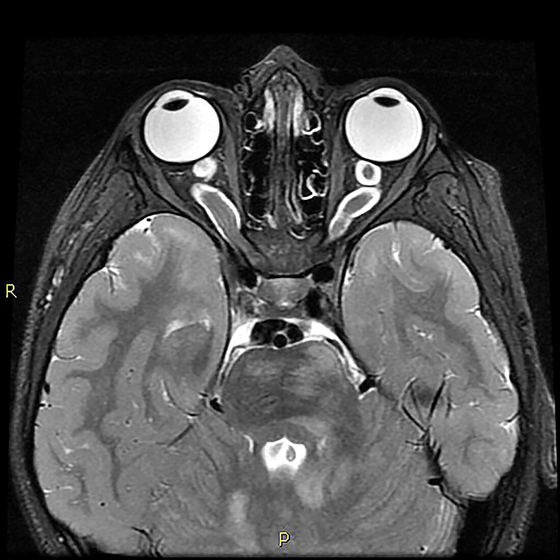
| This axial (cross section) T2 weighted MRI of a 15 year old with known NF1 demonstrates pathologic thickening of the optic nerves within both orbits compatible with optic pathway gliomas. These are common tumours seen in NF1. Also seen are foci of abnormal increased T2 signal within the brainstem and cerebellum which are common findings with NF1. | |
| Licence : | Droits gérés |
| Crédit: | Science Photo Library / Living Art Enterprises |
| Taille de l’image : | 3900 px × 3900 px |
| Model Release : | Non requis |
| Property Release : | Non requis |
| Restrictions : | - |
Prix pour cette image À partir de 45 €
Produit vendu
(Calendrier, Carte postale, Carte de vœux, Impression sur textile, Packaging etc)
À partir de 45 €
Usage commercial
(Affichage, Annonce presse, Annonce TV, Carte, Digital - hors rés. sociaux, Digital - rés. sociaux etc)
À partir de 45 €
Éditorial
(Digital, Journal, Livre, Livre pratique, Magazine, Télévision etc)
À partir de 60 €
Usage non-commercial
(Digital - hors rés. sociaux, Digital - rés. sociaux etc)
À partir de 120 €