Neurofibromatosis type I (NF1), MRI
Numéro d’image : 12636805
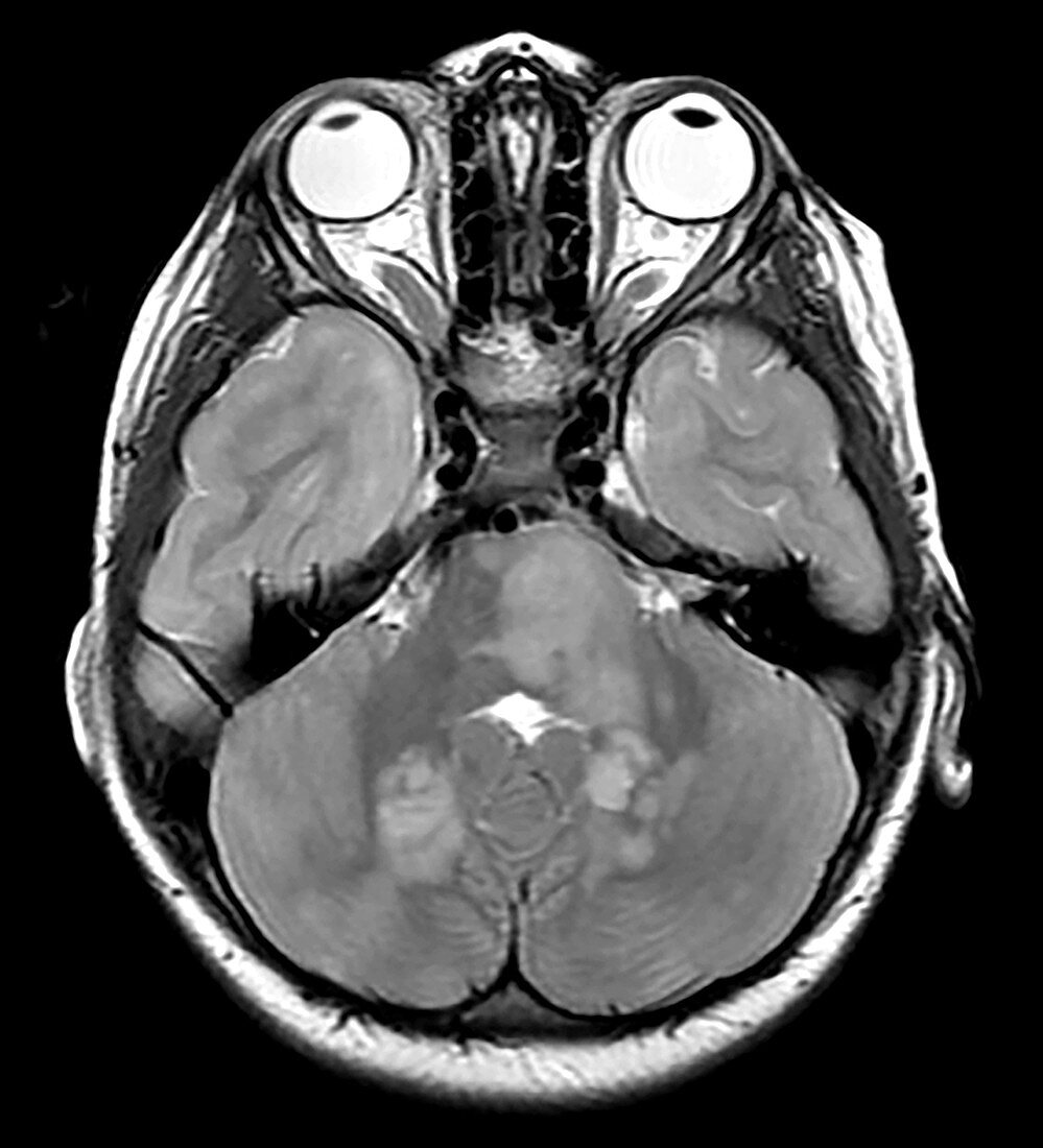
| This axial (cross section) T2 weighted MRI of a 15 year old with known NF1 demonstrates the typical appearance of foci of abnormal increased T2 signal within the brainstem and cerebellum commonly seen in NF1. These foci usually wax and wane and disappear as the subject approaches the age of 20. Also note the enlargement of the optic nerves (right greater than left) with tortuosity and increased CSF in the optic sheaths consistent with optic nerve gliomas. | |
| Licence : | Droits gérés |
| Crédit: | Science Photo Library / Living Art Enterprises |
| Taille de l’image : | 3900 px × 4293 px |
| Model Release : | Non requis |
| Property Release : | Non requis |
| Restrictions : | - |
Prix pour cette image À partir de 45 €
Produit vendu
(Calendrier, Carte postale, Carte de vœux, Impression sur textile, Packaging etc)
À partir de 45 €
Usage commercial
(Affichage, Annonce presse, Annonce TV, Carte, Digital - hors rés. sociaux, Digital - rés. sociaux etc)
À partir de 45 €
Éditorial
(Digital, Journal, Livre, Livre pratique, Magazine, Télévision etc)
À partir de 60 €
Usage non-commercial
(Digital - hors rés. sociaux, Digital - rés. sociaux etc)
À partir de 120 €